Immune System Responses and Tuberculosis Pathogenesis
This content explores innate and adaptive immune proteins, leukocyte functions, and the pathogenesis of Mycobacterium tuberculosis including granuloma formation and the complement cascade.
The human immune system is a sophisticated, multi-layered defense network designed to identify and eliminate invading pathogens. When a pathogen like Mycobacterium tuberculosis enters the body, the immune system does not rely on a single mechanism; instead, it orchestrates a complex interplay between rapid, non-specific innate responses and highly specific, long-lasting adaptive responses. Understanding this interaction is crucial to understanding how certain infections are cleared, how others become latent, and why some pathogens cause chronic disease.
The First Line of Defense: Innate Immunity
Innate immunity serves as the body's immediate response to infection, occurring within hours of colonization. Unlike the adaptive system, the innate response is non-specific, meaning it recognizes general patterns common to many pathogens rather than specific individual strains.
Leukocytes and Inflammation
The primary cellular actors in innate immunity are leukocytes (white blood cells). When a pathogen invades, the body initiates inflammation, a localized immune reaction designed to contain the pathogen and promote its destruction. This process involves several stages of leukocyte recruitment: margination, rolling, firm adhesion, transmigration, and chemotaxis.
Key leukocytes involved include:
Neutrophils: Often the first responders to bacterial infection.
Monocytes and Macrophages: Monocytes circulate in the blood and differentiate into macrophages upon entering tissues. Macrophages are particularly critical in tuberculosis because they are the primary cells responsible for phagocytosis—the process of engulfing and digesting bacteria.
A clinical tool used to assess these cells is the differential leukocyte count, which measures the percentage of each type of white blood cell in a sample. A typical count includes approximately 65% neutrophils, 30% lymphocytes, and 5% monocytes.
Innate Proteins and the Complement System
Beyond cells, the innate response relies on specialized proteins. Cytokines are signaling proteins that promote immune activity; examples include Type I Interferons (IFN-α, IFN-β), TNF-α, and IL-6.
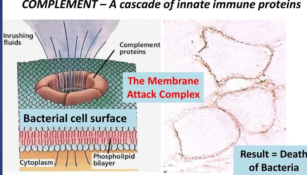
Another vital component is the complement system, a cascade of blood proteins that activates upon infection. This cascade culminates in the formation of the Membrane Attack Complex (MAC), which creates pores in the bacterial cell wall to cause lysis (cell death).
Diagram illustrating the Membrane Attack Complex formation by complement proteins on a bacterial cell surface, leading to bacterial death.
Pathogenesis of Mycobacterium tuberculosis
When Mycobacterium tuberculosis is inhaled via aerosols, it reaches the lungs and is engulfed by alveolar macrophages. However, the pathogen has evolved specific virulence factors to survive this encounter.
Intracellular Survival and Granuloma Formation
Instead of being destroyed, the bacilli can inhibit phagolysosome fusion, a process that would normally allow the macrophage's digestive enzymes to kill the bacteria. This allows the bacteria to replicate intracellularly within the macrophage.
The body typically responds in one of three ways:
The macrophage successfully kills the bacteria.
The macrophage dies, allowing the pathogen to spread.
The formation of a granuloma.
A granuloma is an aggregation of macrophages, matrix, and other immune cells designed to confine the replicating pathogen. A successful immune response results in a non-necrotic granuloma, a structure that is highly dependent on T cells for maintenance. If infected macrophages migrate to local lymph nodes, they may form a Ghon’s focus, which is part of the primary complex.
A diagram illustrating the lifecycle of Mycobacterium tuberculosis from transmission to granuloma formation. This illustrates pathogenesis.
The Specialized Response: Adaptive Immunity
If the innate response cannot clear the infection, the adaptive immune system is activated to provide a specific, targeted attack and establish immunological memory.
Lymphocyte Activation
The bridge between innate and adaptive immunity is provided by dendritic cells and macrophages. These cells traffic M. tuberculosis antigens from the lungs to the lymph nodes. In the lymph node, these antigens activate T lymphocytes and B lymphocytes.
T Lymphocytes: These cells recognize the specific pathogen and proliferate. They produce critical cytokines, such as Type II Interferon (IFN-γ) and IL-2, which coordinate the response and activate macrophages for more efficient killing.
B Lymphocytes: These cells are activated by both the presence of the pathogen and T cell cytokines. Once activated, they produce antibodies (immunoglobulins).
Exam Tip: Note that while antibodies are essential, excessive antibody production can actually lead to inadequate clearance of M. tuberculosis.
The Effector Phase
Once activated, the lymphocytes and their products (antibodies and cytokines) travel back to the site of infection in the lungs via the circulation and lymphatics to enhance the killing of the pathogen.
Clinical Management and Resistance
Diagnosis
The Tuberculin Skin Test (TST) is a common method for detecting TB infection by measuring the induration (swelling) at the site of a PPD injection. The threshold for a positive result depends on the patient's risk profile:
≥5 mm: For immunosuppressed individuals (e.g., HIV-infected) or recent contacts of active TB patients.
≥10 mm: For high-risk groups, such as children, injection drug users, or healthcare workers.
Treatment Regimens
The World Health Organization (WHO) recommends a two-phase treatment approach for new TB patients:
Initial (Intensive) Phase: Typically 2 months of HRZE (Isoniazid, Rifampicin, Pyrazinamide, and Ethambutol) to kill the majority of bacilli.
Continuation Phase: Typically 4 months of HR (Isoniazid and Rifampicin) to eliminate remaining bacilli.
Drug Resistance
A major global health challenge is the rise of drug-resistant strains:
MDR-TB (Multidrug-Resistant TB): Resistant to at least isoniazid and rifampin.
XDR-TB (Extensively drug-resistant TB): Resistant to more than two drugs used in first-line regimens.
These resistance patterns are often driven by premature cessation of therapy, improper medication storage, or transmission in crowded conditions.
Keep reading for free.
Create an account to finish this reading and make it part of your study routine.