Hypertrophic Cardiomyopathy and Sudden Cardiac Death in Athletes
An overview of Hypertrophic Cardiomyopathy (HCM), its genetic basis, pathophysiology, and its role as a leading cause of sudden cardiac death in young athletes. Differentiates HCM from other cardiomyopathies like dilated and restrictive types.
In the world of sports medicine and advanced pathophysiology, few topics are as sobering as the sudden loss of a young, healthy athlete. While we often associate heart disease with aging and long-term lifestyle factors, certain genetic conditions can trigger catastrophic events during moments of peak physical exertion. One of the most critical conditions to understand is Hypertrophic Cardiomyopathy (HCM), a primary driver of sudden cardiac death in young populations.
Understanding Hypertrophic Cardiomyopathy (HCM)
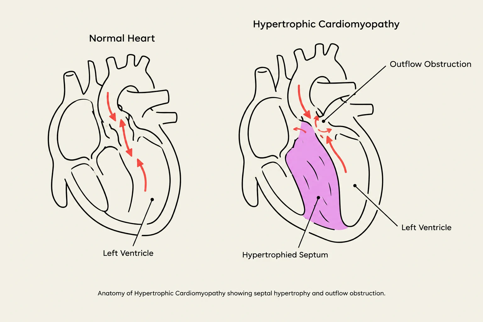
Anatomy of Hypertrophic Cardiomyopathy showing septal hypertrophy and outflow obstruction.
Hypertrophic cardiomyopathy is characterized by an abnormal thickening of the heart muscle, specifically involving the left ventricle. Unlike the thickening seen in patients with chronic hypertension, HCM is an inherited condition where the muscle fibers themselves become enlarged or scarred.
Genetic Basis: Autosomal Dominance
This diagram illustrates the inheritance pattern of an autosomal dominant disorder, showing how alleles are passed from parents to offspring and the resulting genotypes and phenotypes.
HCM is classified as an autosomal dominant genetic disorder. In genetics, "autosomal" means the gene is located on one of the numbered (non-sex) chromosomes, and "dominant" means that an individual only needs to inherit one copy of the mutated gene from one parent to manifest the disease. This high level of heritability explains why the condition can appear across multiple generations of a family.
Pathophysiology and Obstruction
The thickening in HCM is often uneven. A common clinical finding is hypertrophy of the interventricular septum—the wall that separates the left and right ventricles. This uneven thickening can lead to:
Outflow Obstruction: The enlarged septum can physically block the path of blood exiting the heart.
Arrhythmias: The presence of scarred or disorganized muscle fibers can trigger irregular electrical rhythms.
Diastolic Dysfunction: The thickened, stiff walls make it difficult for the ventricle to relax and fill with blood properly.
Clinical Distinction: HCM vs. Hypertensive Hypertrophy
It is vital to distinguish between genetic HCM and acquired left ventricular hypertrophy. In hypertensive hypertrophy, the heart thickens as an adaptation to pumping against high blood pressure; this can often be managed or partially improved by controlling blood pressure. In contrast, genetic HCM is a structural defect of the muscle itself and will not disappear with blood pressure management.
Sudden Cardiac Death (SCD) in Athletes
Sudden Cardiac Death (SCD) refers to a rapid, unexpected death caused by a fatal arrhythmia, typically occurring within an hour of the onset of symptoms. In the context of HCM, SCD often occurs during "vigorous or immediately strenuous exertion".
When an athlete pushes their body to the limit, the increased demand for cardiac output, combined with the structural abnormalities and potential for electrical instability in an HCM patient, creates a "perfect storm" for a fatal event.
Differential Diagnosis: The Cardiomyopathy Spectrum
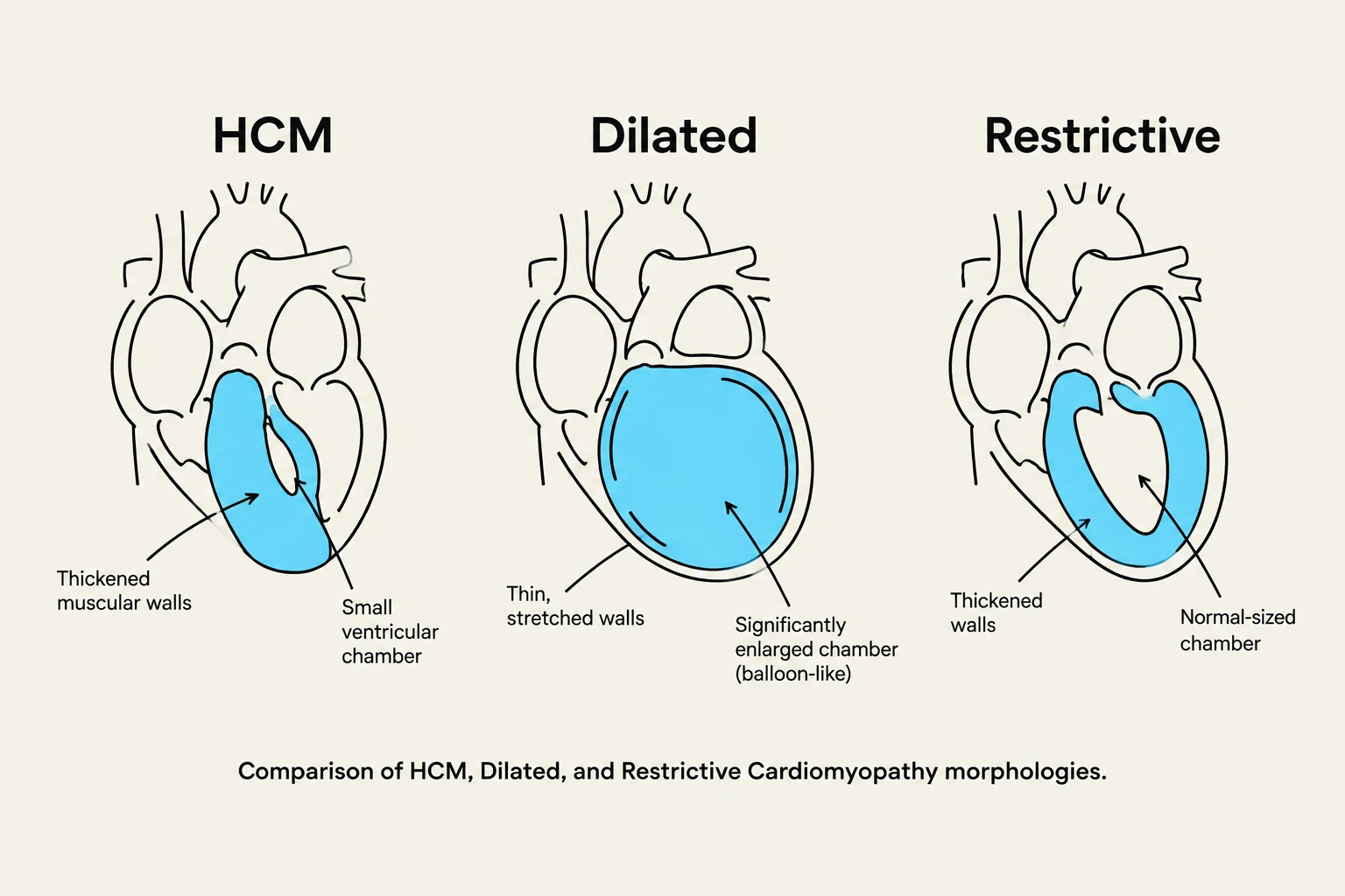
Comparison of HCM, Dilated, and Restrictive Cardiomyopathy morphologies.
To master pathophysiology, one must be able to differentiate HCM from other forms of cardiomyopathy. Understanding these distinctions is essential for clinical accuracy and exam preparation.
1. Dilated Cardiomyopathy
Dilated cardiomyopathy involves the enlargement (dilation) of the left ventricle, leading to a thinning of the heart walls and a decrease in pumping efficiency. Unlike the sudden, exertion-induced death seen in HCM, dilated cardiomyopathy typically presents with progressive symptoms of heart failure, such as:
Chronic fatigue
Dyspnea (shortness of breath)
Fluid retention
2. Restrictive Cardiomyopathy
Restrictive cardiomyopathy is characterized by stiff ventricular walls that resist filling, though the chambers are not necessarily dilated or massively hypertrophied. This is the least common form of cardiomyopathy and is frequently caused by infiltrative systemic diseases in older patients, such as:
Amyloidosis: The buildup of abnormal proteins in the heart tissue.
Sarcoidosis: An inflammatory disease that can cause granulomas to form in the heart muscle.
3. Dystrophic Cardiomyopathy
It is important to note that "dystrophic cardiomyopathy" is not considered one of the standard primary classifications of cardiomyopathy in clinical practice.
Summary Table for Rapid Review
Feature
Hypertrophic (HCM)
Dilated
Restrictive
Primary Change
Thickened, stiff walls (often septal)
Enlarged, thin-walled chambers
Stiff, non-compliant walls
Common Cause
Autosomal dominant genetics
Various (viral, toxic, etc.)
Amyloidosis, Sarcoidosis
Typical Patient
Young athletes / Sudden death
Progressive heart failure
Older adults / Infiltrative disease
Quiz Question
Exam Tip: The "Athlete Scenario"
If you encounter a board-style question describing a young athlete who collapses and dies during a high-intensity practice (like football), your first thought should be Hypertrophic Cardiomyopathy. If the question describes an older patient with progressive shortness of breath and suspected systemic disease, look toward Restrictive Cardiomyopathy.
Keep reading for free.
Create an account to finish this reading and make it part of your study routine.