


The journey from a single cell to a complex, multi-organ organism is one of the most intricate processes in biology. It requires precise hormonal coordination, specialized anatomical structures, and a series of rapid cellular transformations. This article explores the physiological stages of human reproduction, beginning with the female reproductive anatomy and following the development of the embryo through to the formation of a fetus.
The Female Reproductive System and the Uterine Cycle
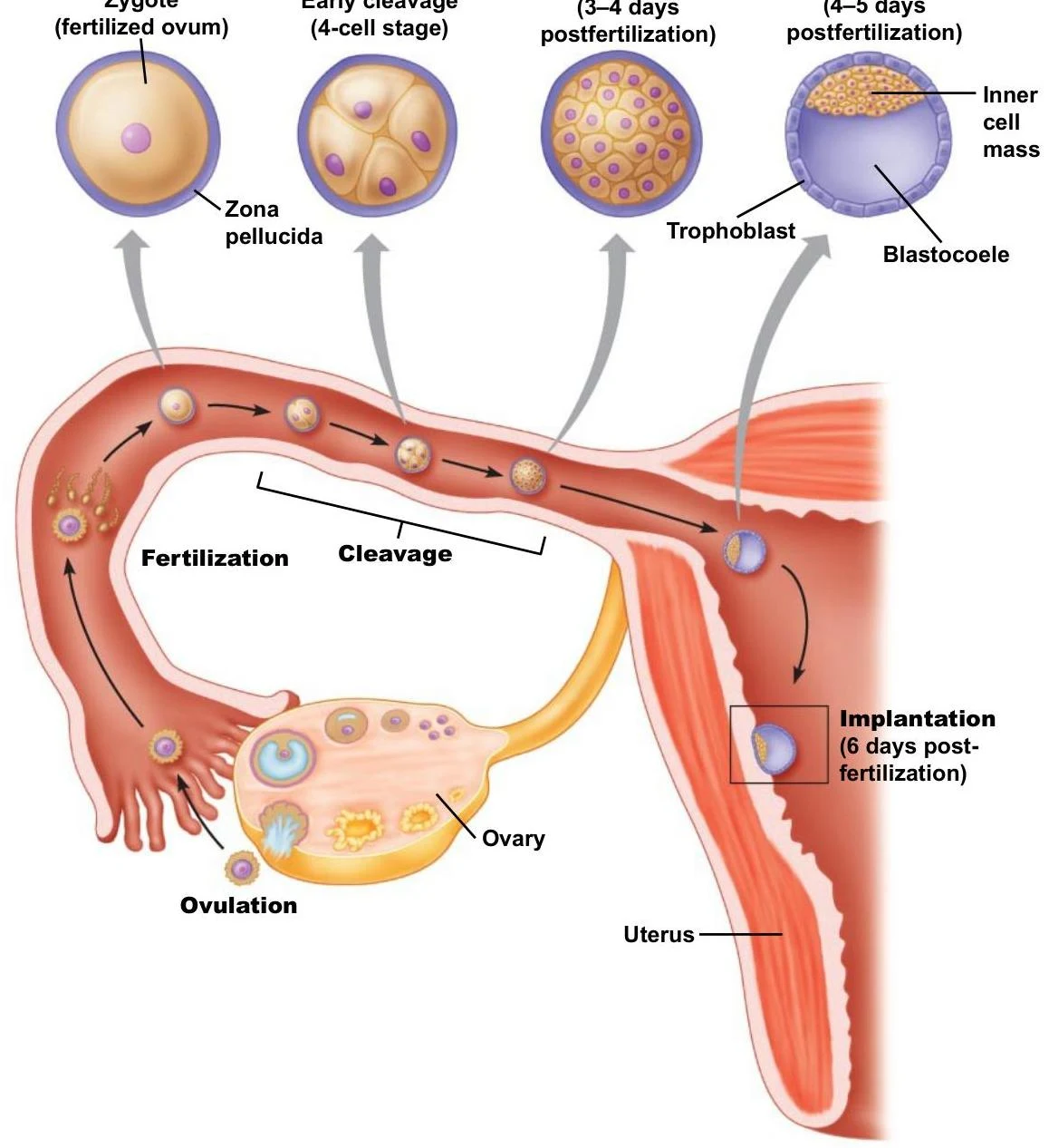
The female reproductive system is designed to facilitate oogenesis (the development of eggs), fertilization, and the support of a developing embryo. Key anatomical structures include the ovaries, the uterine tubes, the uterus, and the vagina—a canal measuring approximately in length that connects the cervix to the external environment.
Within the ovaries, the development of the oocyte is supported by specialized cells known as granulosa cells. These cells play several vital roles: they provide nutrients to the developing oocyte, secrete chemical messengers to support follicle development, and produce estrogens. Notably, granulosa cells also secrete a protein called inhibin.
The uterus undergoes a monthly uterine cycle to prepare for a potential pregnancy. This cycle consists of three distinct phases:
- Menstrual phase: The shedding of the uterine lining.
- Proliferative phase: A critical stage where uterine tissue renews and thickens in preparation for possible implantation.
- Secretory phase: The final preparation stage driven by hormonal changes.


Keep reading for free.
Create an account to finish this reading and make it part of your study routine.
Continue reading freeAlready have an account? Log in
Updated