Purines, such as adenine and guanine, are essential components of life. They serve as the building blocks of DNA and RNA, act as energy carriers (like ATP), and function as critical metabolic regulators and coenzymes. Because these molecules are so vital, the body has evolved complex pathways to both build them from scratch (de novo synthesis) and recycle them (the salvage pathway). However, when the breakdown of these molecules goes awry, the resulting metabolic byproducts can lead to severe clinical conditions ranging from painful joint inflammation to profound neurological impairment.
Purine Biodegradation and Uric Acid Production
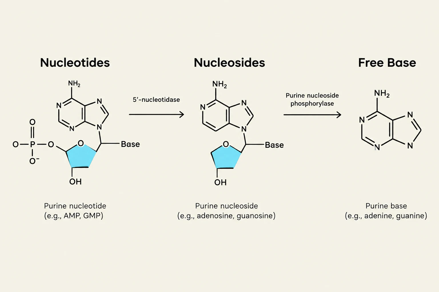
When purine nucleotides are no longer needed, they undergo a degradation process to be excreted from the body. This catabolic pathway involves several steps where nucleotidases and nucleosidases first release the free purine bases from their sugar and phosphate components.
The central goal of human purine biodegradation is the production of uric acid. The process follows a specific enzymatic route:
Free bases (like hypoxanthine and guanine) are converted into xanthine via enzymes such as guanine deaminase.
The enzyme xanthine oxidase then acts on these intermediates. It is a versatile enzyme capable of oxidizing two different sites on the purine ring system.
Xanthine oxidase converts xanthine into the final metabolic product: uric acid.
This diagram illustrates the biochemical breakdown of purine nucleotides into nucleosides and then into free purine bases, catalyzed by specific enzymes.
Clinical Note: Unlike pyrimidine degradation, which produces highly water-soluble products like β-alanine and CO2, purine degradation produces uric acid, which has relatively low solubility in biological fluids.
Metabolic Defects: Lesch-Nyhan Syndrome
One of the most striking examples of purine metabolic dysfunction is Lesch-Nyhan Syndrome. This condition is caused by a severe deficiency in the enzyme HGPRT (hypoxanthine-guanine phosphoribosyltransferase).
Under normal conditions, HGPRT is a key player in the salvage pathway, which recycles free bases (hypoxanthine and guanine) by attaching them back to PRPP (5-phosphoribosyl-1-pyrophosphate) to reform nucleotides. In Lesch-Nyhan Syndrome, the lack of HGPRT activity leads to a cascade of metabolic failures:
PRPP Accumulation: Because the salvage pathway is blocked, PRPP is not consumed and builds up in the cell.
Increased De Novo Synthesis: High levels of PRPP act as a potent activator for the de novo purine synthesis pathway.
Uric Acid Overproduction: The combination of decreased purine recycling and increased de novo synthesis results in a massive excess of purines being funneled into the degradation pathway, creating enormous amounts of uric acid.
This syndrome is an X-linked recessive trait, occurring primarily in males, and is characterized by gouty arthritis and severe neurological malfunctions, including mental retardation, aggressiveness, and self-mutilation.
Hyperuricemia and Gout
When the concentration of uric acid in the blood exceeds normal physiological limits, the condition is known as hyperuricemia. Normal plasma levels are typically around 2.4–6 mg/dL for females and 3.4–7 mg/dL for males; a level greater than 7.0 mg/dL is considered hyperuricemic.
Hyperuricemia can be categorized into two types:
Primary: An innate defect in how the body metabolizes purines or excretes uric acid.
Secondary: Caused by external factors such as diet, medications, or other medical conditions.
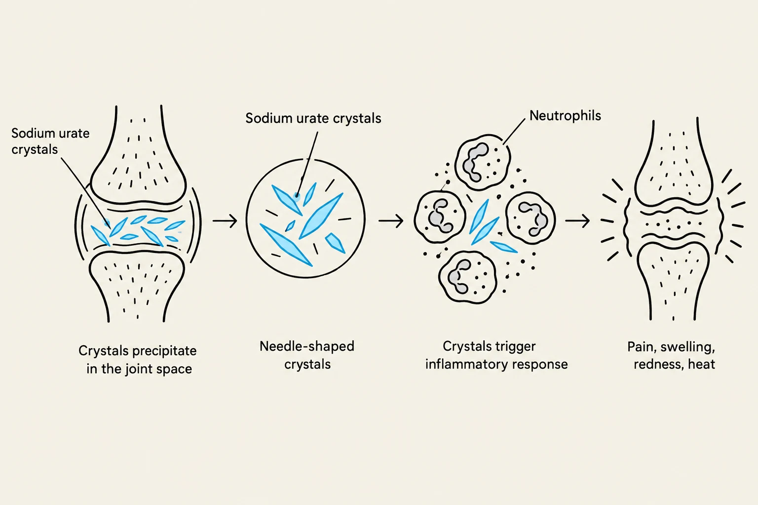
If uric acid levels remain high, the acid can precipitate out of the solution, forming sodium urate crystals (also known as tophi). When these needle-shaped crystals deposit in the joints, they trigger intense inflammation and pain, a condition known as gout (or gouty arthritis).
This diagram illustrates the process by which sodium urate crystals precipitate in a joint space, triggering an inflammatory response characterized by pain, swelling, redness, and heat.
Diagnosis and Treatment of Gout
To diagnose gout, clinicians often perform an aspiration of synovial fluid from an affected joint. Under polarized light microscopy, the presence of needle-shaped monosodium urate crystals confirms the diagnosis.
Treatment strategies include:
Allopurinol: A potent inhibitor of xanthine oxidase. It is an analog of hypoxanthine that prevents the conversion of xanthine to uric acid. This results in an accumulation of hypoxanthine and xanthine, which are much more soluble than uric acid.
Colchicine: Used to reduce inflammation.
Uricosuric agents (e.g., probenecid): Used to increase the renal excretion of uric acid.
Dietary Management: Avoiding high-purine foods (red meats, organ meats, certain vegetables like spinach or mushrooms) and limiting alcohol and caffeine.
Kidney Stones and SCID
High concentrations of uric acid do not only affect the joints. When uric acid is present in high levels in the blood, it can precipitate as a salt within the kidneys, forming kidney stones. These stones can lead to significant pain, infection, and potential kidney damage.
Finally, defects in the purine pathway can impact the immune system. A deficiency in the enzyme adenosine deaminase (ADA) leads to an accumulation of adenosine and deoxyadenosine. The resulting increase in dATP levels inhibits the enzyme ribonucleotide reductase. Because ribonucleotide reductase is essential for producing the deoxyribonucleotides needed for DNA synthesis, cells (particularly lymphocytes) cannot divide. This manifests as Severe Combined Immunodeficiency (SCID), a condition characterized by a critical decrease in both T cells and B cells.
Exam Tip: Remember the distinction between the two major purine pathways. The Salvage Pathway recycles bases to save energy, while the De Novo Pathway builds nucleotides from scratch. Defects in the salvage pathway (like HGPRT deficiency) often lead to an increase in the de novo pathway, ultimately causing hyperuricemia.
Keep reading for free.
Create an account to finish this reading and make it part of your study routine.