Human Leukocyte Antigens (HLA) and Immune Response
An overview of Human Leukocyte Antigens (HLA), their role in distinguishing self from non-self, and their critical importance in immunology and organ transplantation.
In the complex landscape of the human immune system, one of the most critical challenges is distinguishing between the body's own healthy cells and foreign invaders like bacteria, viruses, or transplanted tissue. This distinction is governed by a specialized group of proteins known as the Major Histocompatibility Complex (MHC). In humans, these proteins are specifically referred to as Human Leukocyte Antigens (HLA). Understanding the genetic variation of HLA is not only fundamental to immunology but is also the cornerstone of modern transplant medicine.
What are HLA Proteins?
The HLA system consists of a group of genes located on chromosome 6 that encode cell-surface glycoproteins. These proteins act as "identification tags" for cells. Their primary function is to present peptide fragments—small pieces of proteins—to T cells. Depending on the type of HLA molecule, these peptides can be derived from either internal cellular processes (endogenous) or from external sources that have been engulfed by the cell (exogenous).
The HLA system is categorized into two main classes:
Class I HLA: Found on almost all nucleated cells in the body. These molecules present peptides to CD8+ cytotoxic T cells. They are essential for signaling that a cell is infected by a virus or has become cancerous.
Class II HLA: Found primarily on specialized "professional" antigen-presenting cells (APCs), such as macrophages, B cells, and dendritic cells. These molecules present peptides to CD4+ helper T cells to coordinate a broader immune response.
Diagrams illustrating the structure and function of Class I and Class II HLA molecules, including their peptide binding characteristics and cellular locations.
Exam Tip: Remember the "Rule of 8." Class I molecules interact with CD8+ T cells (1×8=8), and Class II molecules interact with CD4+ T cells (2×4=8). This is a quick way to keep the cell types straight during a multiple-choice exam.
The Power of Genetic Variation: Polymorphism and Polygeny
What makes the HLA system unique compared to most other genetic systems is its extreme level of variation. This variation is driven by two key genetic principles: polygeny and polymorphism.
Polygeny
Polygeny refers to the fact that humans have multiple different genes that encode for similar types of proteins. For example, we do not just have one "HLA Class I" gene; we have several (HLA-A, HLA-B, and HLA-C). This allows for a wider variety of peptide presentation.
Polymorphism
Polymorphism refers to the existence of many different alleles (versions) of these genes within the human population. While most genes have only a few variations, HLA genes are among the most polymorphic in the entire human genome. This means that if you were to pick two random people from the street, the likelihood of them having identical HLA profiles is incredibly low.
The biological advantage of this extreme variation is evolutionary: it ensures that even if a new pathogen evolves to "hide" from one person's HLA molecules, it is unlikely to be able to hide from the HLA molecules of the entire population.
This illustration compares low genetic diversity with high HLA polymorphism, showing how a population with greater HLA diversity has a broader ability to recognize different non-self antigens.
HLA and Organ Transplantation
The very feature that protects the human species from disease—extreme genetic variation—is the greatest obstacle in organ transplantation. When a donor organ is transplanted into a recipient, the recipient's immune system scans the cells of the new organ.
If the donor's HLA molecules are significantly different from the recipient's, the recipient's T cells will recognize them as "non-self" or foreign. This recognition triggers an immune attack, leading to transplant rejection.
Types of Rejection
Rejection can be categorized based on the timing and the mechanism of the immune response:
Hyperacute Rejection: Occurs within minutes to hours. This is usually caused by pre-existing antibodies in the recipient that recognize the donor's HLA, leading to immediate destruction of the organ's blood vessels.
Acute Rejection: Occurs within days to weeks or months. This is primarily mediated by T cells that recognize the mismatched HLA molecules as foreign antigens.
Chronic Rejection: Occurs over months or years. This involves a slow, progressive immune-mediated damage that eventually leads to organ failure.
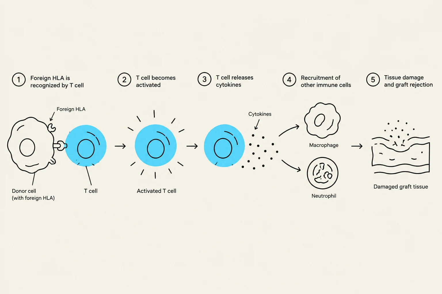
This diagram illustrates the process of graft rejection, where a T cell recognizes foreign HLA on a donor cell, becomes activated, releases cytokines, recruits other immune cells like macrophages and neutrophils, leading to tissue damage and rejection of the graft.
Clinical Implications: Matching and Immunosuppression
To minimize the risk of rejection, clinicians perform HLA typing on both the donor and the recipient before a transplant is attempted. The goal is to find a "match"—a donor whose HLA alleles are as close as possible to those of the recipient. This is why siblings are often the best candidates for organ donation, as they are more likely to share HLA alleles.
Even with a good match, the immune system is highly sensitive. Therefore, transplant recipients must undergo lifelong immunosuppressive therapy. These drugs work by dampening the activity of T cells and other immune components, preventing them from mounting a full-scale attack against the "foreign" HLA molecules of the transplanted organ.
While immunosuppression is necessary to save the organ, it comes with a trade-off: the patient becomes more susceptible to opportunistic infections and certain types of cancer because their overall immune surveillance is diminished.
Keep reading for free.
Create an account to finish this reading and make it part of your study routine.