An in-depth exploration of the human foot's structure, including its three main regions (forefoot, midfoot, rearfoot), ossification process, and the biomechanics of pronation and supination. It also covers vascular supply, venous return mechanisms, and the sensory and motor innervation crucial for foot function and bal
The human foot is a marvel of biological engineering, serving as both a shock absorber and a high-performance lever. It is a complex structure divided into three primary regions: the forefoot (metatarsals and phalanges), the midfoot (navicular, cuboid, and cuneiforms), and the rearfoot (talus and calcaneus). To function effectively, the foot relies on a sophisticated integration of bone development, triplanar joint mechanics, a robust vascular network, and precise neural control. Understanding these systems is essential for grasping how the foot adapts to uneven terrain and how clinical interventions, such as orthotics, influence the entire kinetic chain.
Development and Ossification
Unlike many other bones, the foot is predominantly cartilaginous at birth. Its development follows a specific ossification sequence that dictates how a child’s foot appears and functions over time.
Primary Ossification: The calcaneus, talus, and cuboid begin the ossification process around birth or during late fetal development.
Late Ossification: The navicular is typically the last of the tarsal bones to ossify, a process that usually occurs between ages 2 and 5.
Fusion: The secondary ossification centers (epiphyses) of the metatarsals and phalanges do not fuse until adolescence, typically between ages 14 and 18.
Because of this high degree of ligamentous laxity and the presence of a prominent subcutaneous fat pad, infants and toddlers often present with a "flexible flatfoot" appearance. The visible medial longitudinal arch typically begins to establish itself between ages 4 and 6 as intrinsic muscle strength improves and the fat pad resolves.
Biomechanics: The Subtalar Joint and Posture
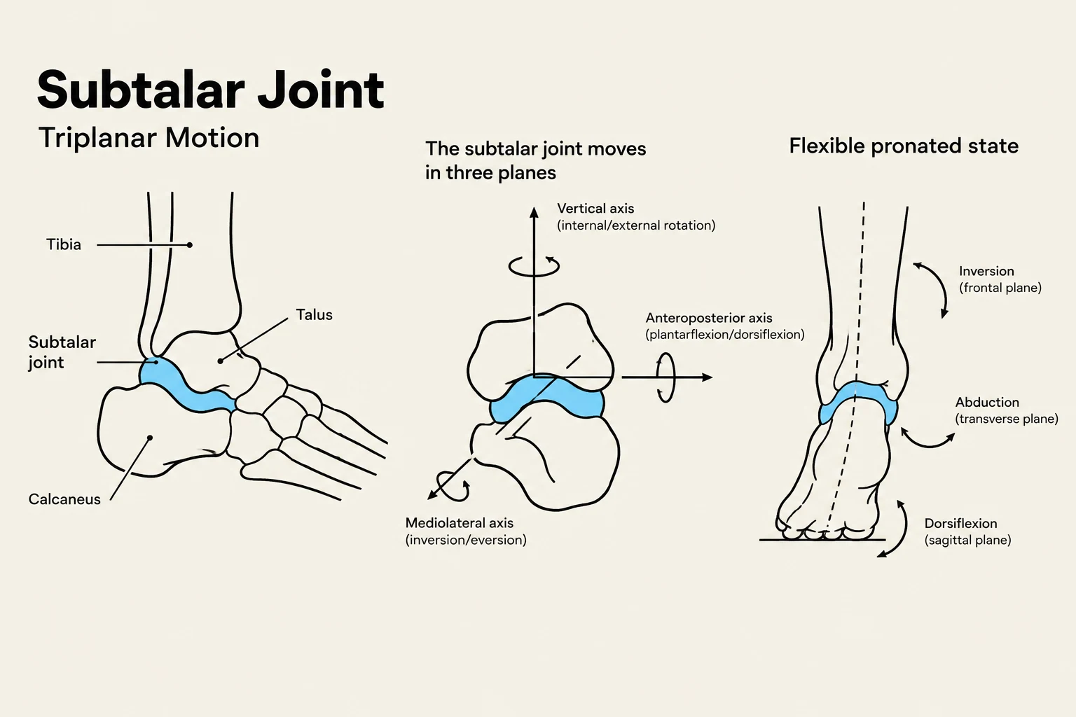
The subtalar joint (STJ), or talocalcaneal joint, acts as a mechanical "torque converter" between the lower leg and the ground. It does not function as a simple hinge; instead, it operates on an oblique axis, allowing for triplanar motion.
Pronation vs. Supination
The STJ transitions between two critical states during the gait cycle to balance flexibility and stability:
Pronation (The Mobile Adapter): Occurring during the early stance phase (heel strike to midstance), pronation involves a combination of eversion, abduction, and dorsiflexion. This movement "unlocks" the midtarsal joint, allowing the foot to become a flexible adapter that absorbs ground reaction forces and conforms to uneven terrain.
Supination (The Rigid Lever): During the propulsive phase (midstance to toe-off), the STJ undergoes supination—a combination of inversion, adduction, and plantarflexion. This action "locks" the midfoot and forefoot bones together, transforming the foot into a rigid lever capable of efficient forward propulsion.
Diagram illustrating the subtalar joint and its triplanar motion, including inversion, abduction, and dorsiflexion.
Foot Types and Posture
Foot posture is often classified by the height of the arches:
Normal (Rectus): Features a neutral arch and even load distribution.
Pes Planus (Flat Foot): Characterized by a collapsed or low medial longitudinal arch. This can be flexible or rigid (e.g., tarsal coalition).
Pes Cavus (High Arch): Features a high arch with reduced contact area, often resulting in lateral loading of the foot.
Clinicians often use the Foot Posture Index (FPI-6) to score static posture, ranging from -12 (highly supinated) to +12 (highly pronated).
Exam Tip: Remember that the STJ axis of rotation is highly individualized. Because of the "mitered hinge" model, any orthotic adjustment at the foot level will alter the proximal torque and forces transmitted up the entire kinetic chain.
Vascular and Venous Physiology
The foot requires a high metabolic supply to support the intense work of the intrinsic and extrinsic muscles. This is achieved through a branched network of anastomoses (interconnections).
Arterial Supply
Dorsal Supply: The anterior tibial artery becomes the dorsalis pedis artery, supplying the dorsum of the foot via the arcuate artery.
Plantar Supply: The posterior tibial artery divides into the medial plantar and lateral plantar arteries. The lateral plantar artery is particularly important as it helps form the deep plantar arch.
Venous Return and the Musculovenous Pump
Since blood must travel against gravity to return to the heart, the foot relies on two primary mechanisms:
Venous Valves: One-way bicuspid valves in the veins ensure unidirectional flow and prevent retrograde flow (reflux).
Musculovenous Pump: The contraction of the gastrocnemius and soleus muscles compresses the deep veins, forcing blood upward.
Innervation: Sensory and Motor Control
The nervous system provides the continuous proprioceptive feedback and motor commands necessary for balance and movement.
Sensory Innervation (Cutaneous Sensation)
Different nerves cover specific territories of the foot:
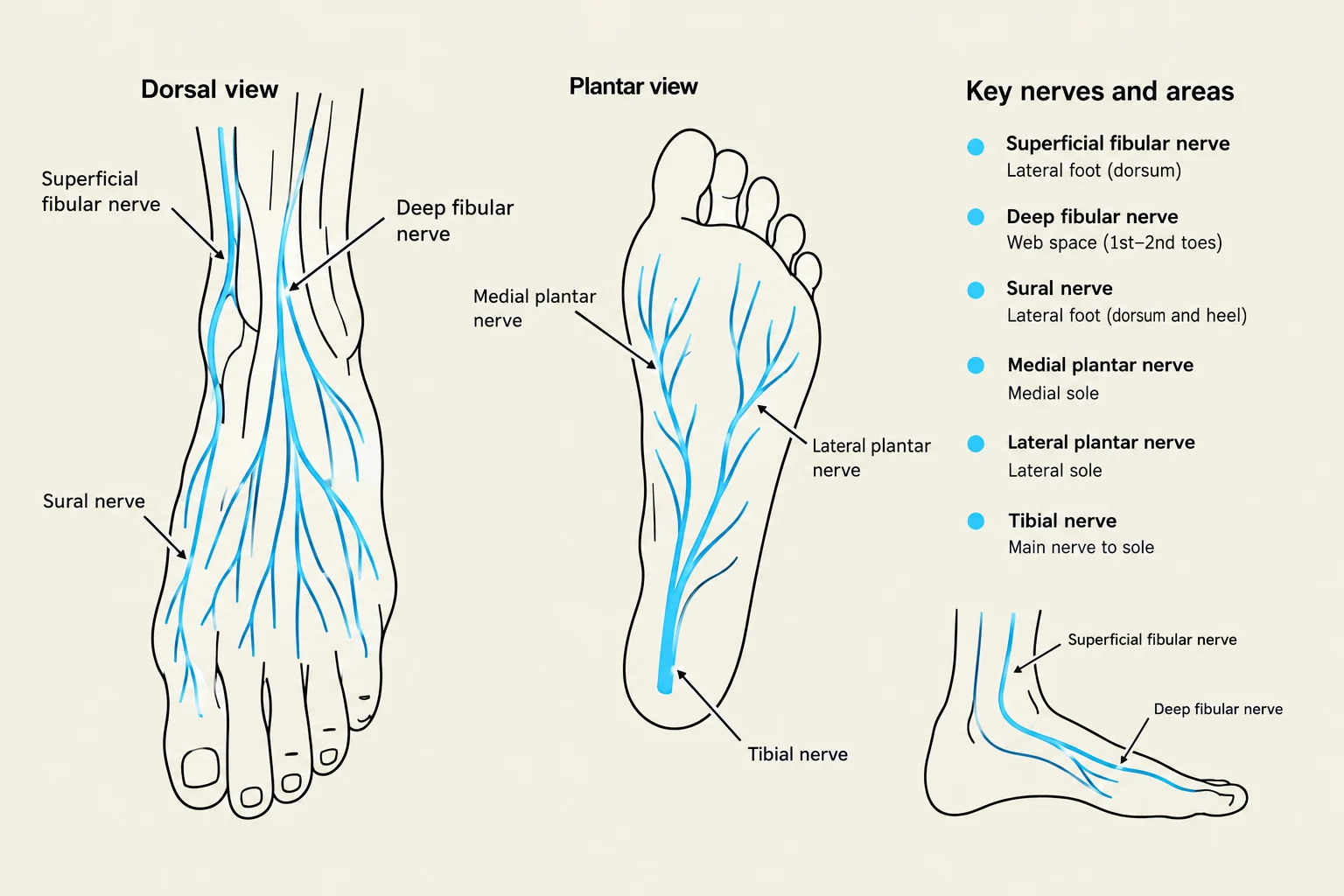
Superficial Fibular Nerve: Supplies most of the dorsum (top) of the foot.
Deep Fibular Nerve: Supplies the first web space (between the great toe and second toe).
Sural Nerve: Supplies the lateral edge and lateral heel.
Plantar Nerves: The medial and lateral plantar nerves (branches of the tibial nerve) supply the sole of the foot.
Motor Innervation
Motor control is divided between extrinsic muscles (located in the leg) and intrinsic muscles (located within the foot):
Extrinsic Muscles: Controlled by the deep fibular nerve (anterior compartment/dorsiflexors), the superficial fibular nerve (lateral compartment/everters), and the tibial nerve (posterior compartment/plantarflexors).
Intrinsic Muscles: Nearly all intrinsic muscles of the sole are innervated by the tibial nerve via its medial and lateral plantar branches.
Diagrams illustrating the dorsal and plantar views of the nerves in the human foot, along with a key identifying specific nerves and their corresponding areas.
Nerve
Primary Motor Action
Primary Sensory Territory
Deep Fibular
Dorsiflexion & Toe Extension
First web space
Superficial Fibular
Eversion
Dorsum of the foot
Tibial (Plantar branches)
Toe Flexion & Arch Support
Sole of the foot
Clinical Note: In-toeing in children can arise from various levels: the foot (metatarsus adductus), the tibia (internal tibial torsion), or the femur (femoral anteversion). Most cases resolve spontaneously, but they significantly impact the foot progression angle.
Keep reading for free.
Create an account to finish this reading and make it part of your study routine.