Human Embryonic Development and the Four Primary Tissue Types
An overview of human embryonic development, from fertilization to the formation of germ layers, and a detailed explanation of the four primary tissue types: epithelial, connective, muscle, and nervous tissue.
The journey from a single cell to a complex, multi-layered human organism is one of nature's most intricate processes. This transformation involves precise cell divisions, the specialization of cells into specific roles, and the physical reshaping of the body. To understand how a human being is formed, we must look at two interconnected biological stages: the early embryonic development that builds the foundation of life, and the formation of the four primary tissue types that allow organs to function.
From Zygote to Blastocyst: The Early Stages
The process of life begins with fertilization, resulting in a zygote, a single diploid cell. This zygote does not remain a single cell for long; it immediately begins to divide through a process called mitosis. During mitosis, the cell replicates its DNA and divides to create identical daughter cells.
The division follows a predictable sequence: 1 cell becomes 2, then 4, then 8, and eventually reaches the 16-cell stage, known as the morula. As division continues, the cluster of cells undergoes further organization to become a blastocyst (the mammalian version of a blastula) around day 4–5 after fertilization.
The blastocyst is a hollow, spherical structure with distinct specialized regions:
Trophoblast: The outer layer of cells that will eventually develop into the placenta.
Inner Cell Mass (Embryoblast): The internal cluster of cells that serves as the source of the embryo and fetus.
Blastocoel: The internal fluid-filled cavity that becomes the yolk sac.
Once the blastocyst reaches the uterine endometrium, it undergoes implantation. This is a critical step for the pregnancy to proceed.
Gastrulation and the Formation of Germ Layers
Around day 14, the embryo undergoes a transformative process called gastrulation, marked by the appearance of the primitive streak. During this stage, the cells organize into three foundational germ layers. These layers are the "blueprints" from which all specific tissues and organs will eventually emerge.
As the embryo develops, two fundamental biological processes drive its complexity:
Differentiation: This is the specialization of cells to assume specific forms and functions. This process relies on stem cells. Pluripotent cells are highly versatile and can differentiate into any cell type in the human body, whereas adult stem cells are more limited, capable of only producing specific cell types within a particular organ or tissue.
Morphogenesis: This refers to the physical change in the embryo's shape and the organized functional arrangement of its parts.
As the third embryonic week progresses, the embryo begins to form vital structures such as the neural tube, which is the precursor to the central nervous system.
Exam Tip: Remember the distinction between gestational age and fetal age. As the embryo develops into a fetus, the biological age of the organism (fetal age) may differ from the time elapsed since the last menstrual period (gestational age).
The Four General Types of Tissues
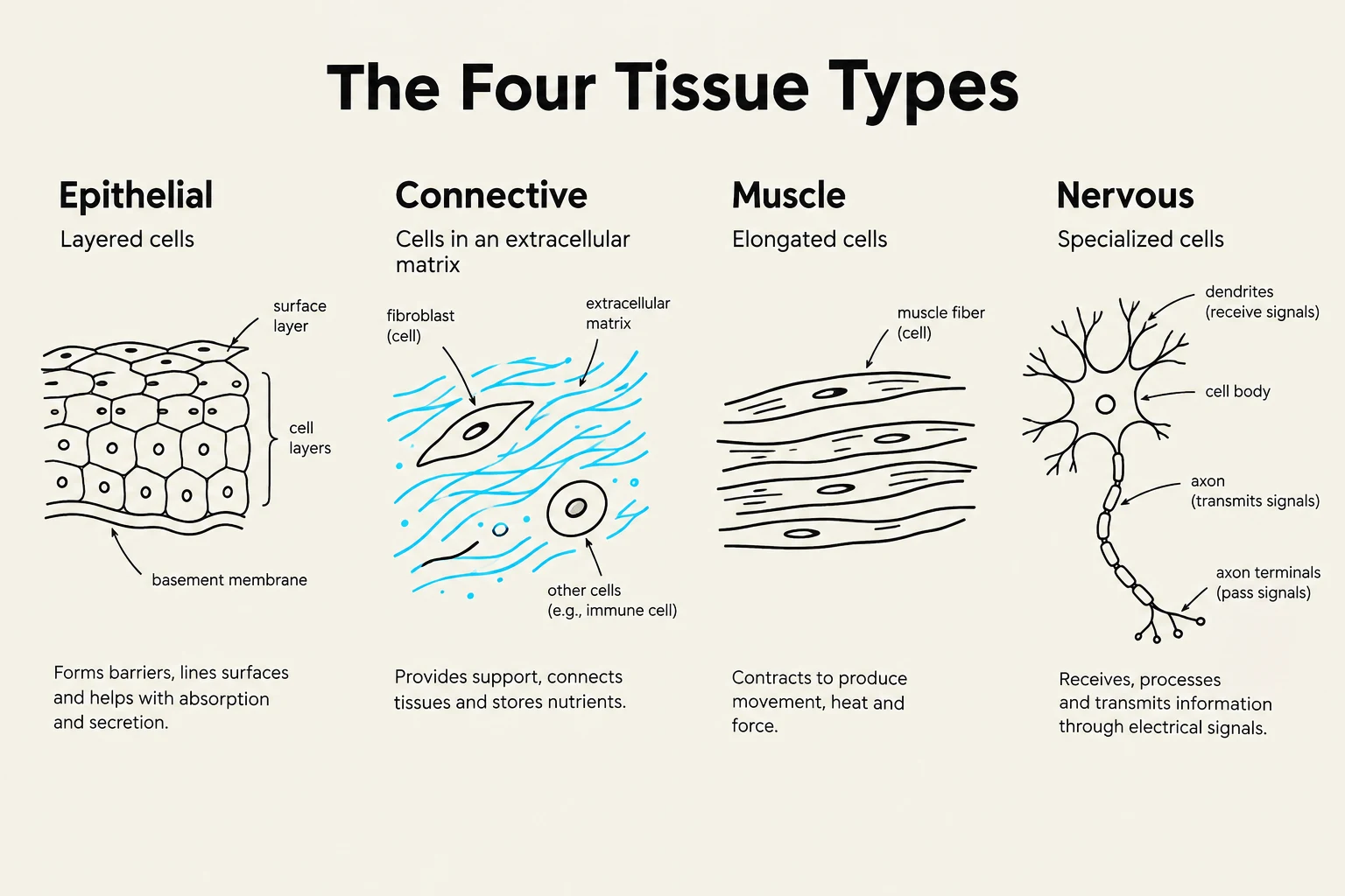
As the germ layers differentiate, they form the four primary types of tissues that compose the human body. A tissue is defined as a collection of cells of the same type that work together to perform a common function.
1. Epithelial Tissue: The Body's Coverings
Epithelial tissues cover body surfaces and line cavities and tubes. They are classified by their shape (squamous, cuboidal, or columnar) and their organization (simple, which is one layer, or stratified, which is multiple layers).
Simple Squamous: A single layer of flat cells used for diffusion and filtration, such as in the air sacs of the lungs.
Simple Cuboidal/Columnar: Often found in glands and the gut, these are specialized for secretion and absorption.
Stratified Epithelium: Multiple layers designed for protection, such as the skin.
Transitional Epithelium: Unique cells that can change shape to allow organs like the bladder to stretch.
Additionally, epithelial tissue forms glands, which are categorized as exocrine (secreting into ducts, like sweat glands) or endocrine (secreting hormones directly into the bloodstream).
2. Connective Tissue: Support and Binding
Connective tissue is the most abundant tissue type and serves to connect, support, and anchor body parts. It is characterized by an extracellular matrix (EM), which consists of ground substance and fiber-like proteins like collagen and elastin.
The three main categories are:
Fibrous (Soft) Connective Tissue: Includes loose connective tissue (like adipose/fat) and dense connective tissue (like tendons and ligaments).
Supportive Connective Tissue: Includes cartilage (hyaline, elastic, and fibrocartilage) and bone.
Fluid Connective Tissue: Includes blood (composed of plasma and formed elements like RBCs and WBCs) and lymph.
Diagram illustrating the four main types of human tissue: epithelial, connective, muscle, and nervous tissue, along with their characteristics and functions.
3. Muscle Tissue: Movement and Heat
Muscle tissue is specialized for movement and the production of heat. It is composed of muscle fibers (myocytes) containing protein filaments called actin and myosin.
There are three distinct types:
Skeletal Muscle: Striated, multi-nucleated, and under voluntary control; it is attached to bones via tendons.
Smooth Muscle: Non-striated, spindle-shaped, and involuntary; found in the walls of hollow organs like the stomach.
Cardiac Muscle: Striated, branched, and involuntary; found exclusively in the heart and characterized by intercalated disks.
4. Nervous Tissue: Communication
Nervous tissue is responsible for detecting, processing, and coordinating information through sensory input, integration, and motor output. It consists of two main cell types:
Neurons: The functional units that carry messages via dendrites (receiving information) and axons (sending information).
Neuroglia (Glial cells): The support cells that make up about 90% of the nervous system. They provide nutrients, physical support, and protection to neurons.
Study Note: While neurons carry the electrical signals, they do not actually touch. Communication occurs at a synapse, where chemicals called neurotransmitters are released to bridge the gap between cells.
By the end of these processes, the organized interaction of tissues and germ layers allows for the development of complex organ systems, ensuring the body can maintain homeostasis and support life.
Keep reading for free.
Create an account to finish this reading and make it part of your study routine.