


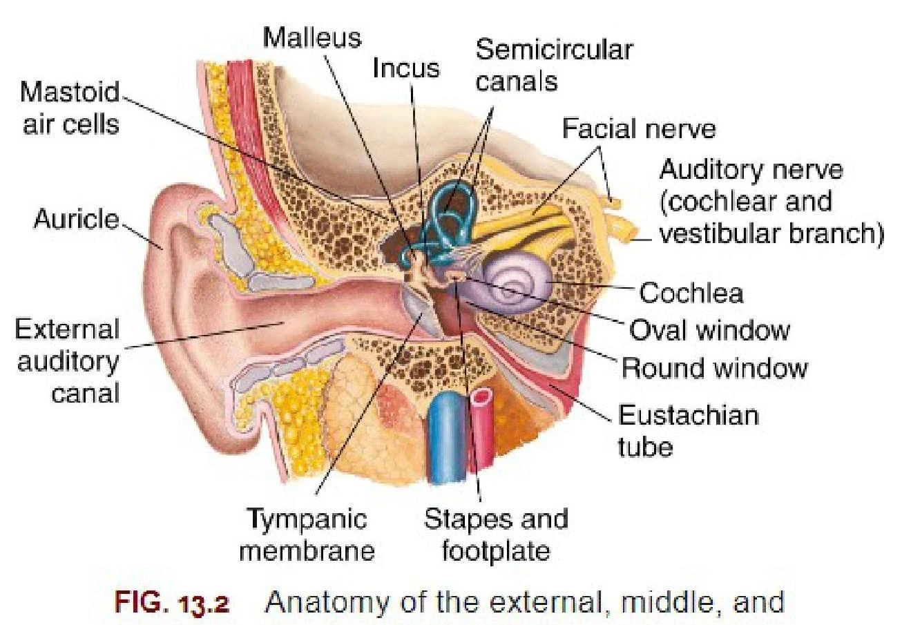
The human ear is a sophisticated sensory organ responsible for two critical functions: hearing and maintaining equilibrium (balance). To achieve these tasks, the ear is organized into three distinct anatomical regions: the external ear, the middle ear, and the inner ear. Each region serves as a specialized link in a chain that converts mechanical sound waves into electrical signals that the brain can interpret.
The External Ear: Sound Collection
The journey of sound begins with the external ear, which acts as a funnel to capture acoustic energy from the environment. The most visible part of this system is the auricle (also known as the pinna).
The auricle is a complex cartilaginous structure with several distinct landmarks that help direct sound into the ear canal. Key features include:
- Helix: The prominent outer rim of the auricle.
- Antihelix: The curved ridge located just inside the helix.
- Triangular fossa: The depression located between the helix and the antihelix.
- Concha: The deep, bowl-like hollow that leads into the ear canal.
- Antitragus: A small cartilaginous projection opposite the tragus.
- Lobule: The fleshy, non-cartilaginous lower part of the ear.
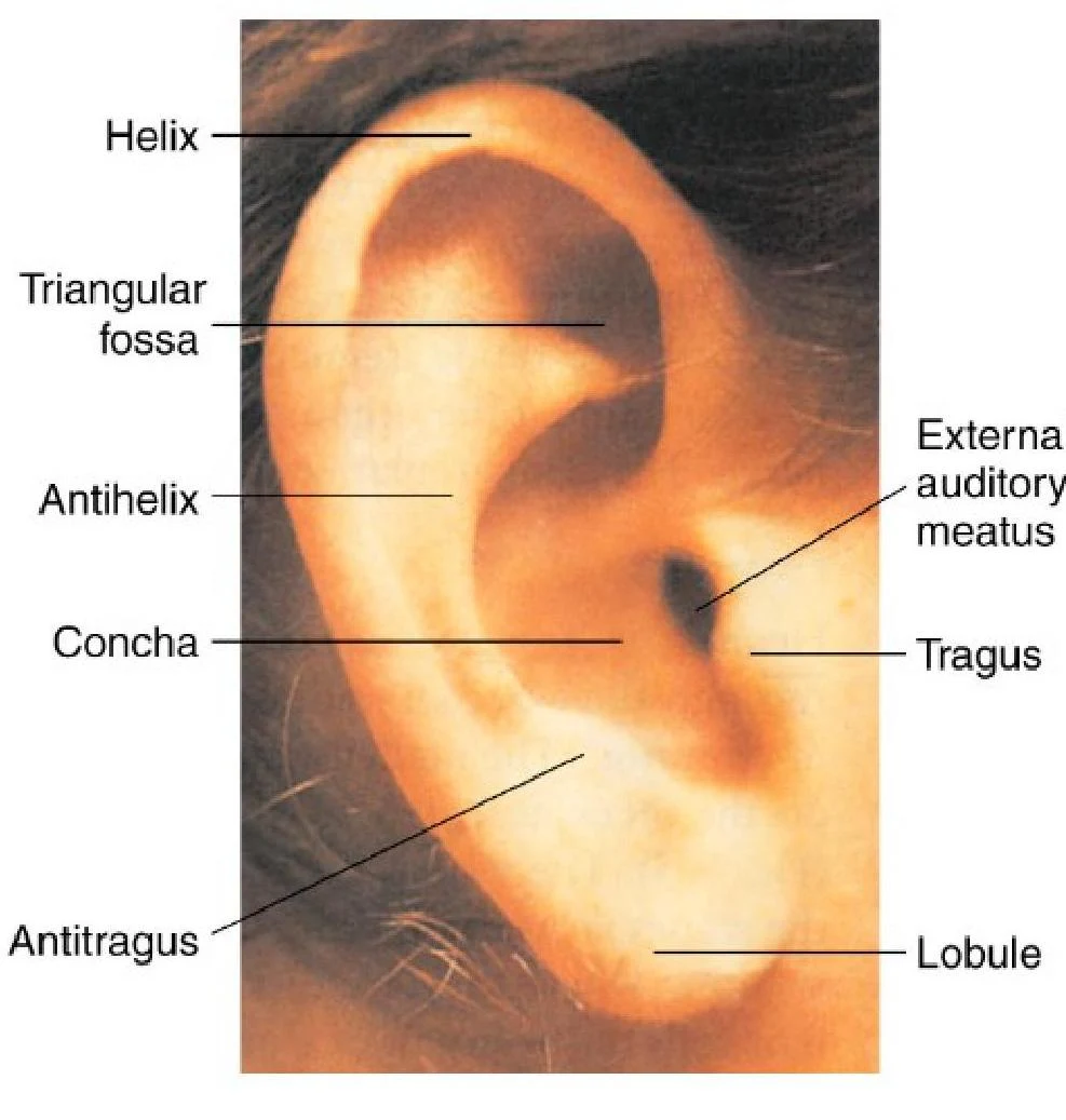
Once collected by the auricle, sound waves travel through the external auditory canal. This canal serves as a passageway leading directly to the middle ear.
Clinical Note: Acute Otitis Externa Inflammation of the external ear canal, often called "swimmer's ear," can cause severe pain (otalgia) when the pinna is manipulated. Common signs include edema, erythema, and sometimes yellow-greenish drainage.
Keep reading for free.
Create an account to finish this reading and make it part of your study routine.
Continue reading freeAlready have an account? Log in
Updated