


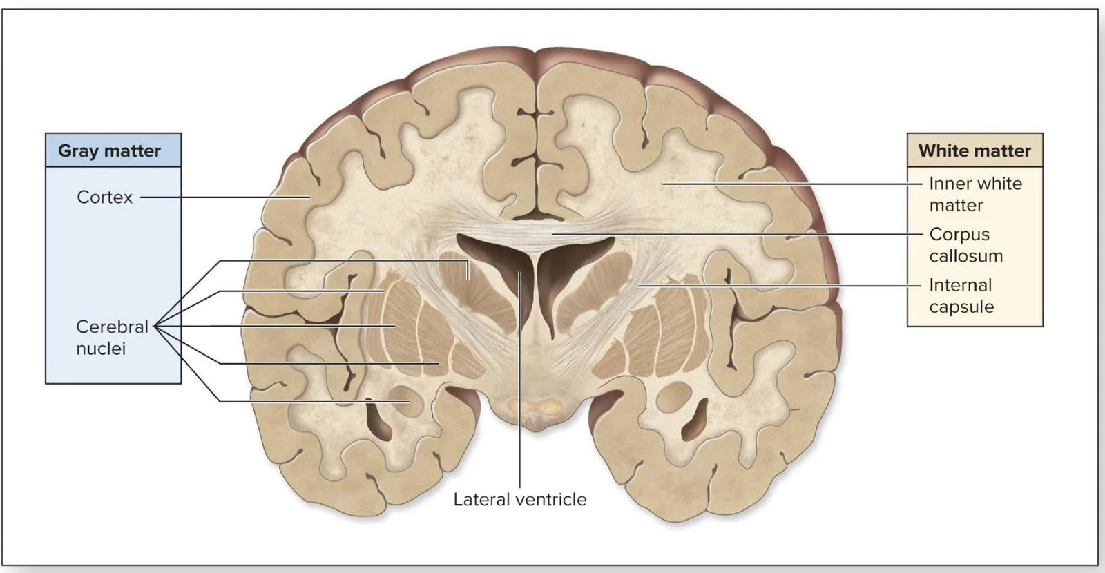
The human brain is one of the most complex organs in the body, characterized by a highly specific structural organization that emerges during early development. To understand how the adult brain functions, one must first look back at its embryonic origins and the way neural tissues organize into the functional layers we observe in adulthood. This article explores the progression from embryonic vesicles to mature brain structures and the fundamental organization of neural tissue.
Embryonic Development and Brain Vesicles
The development of the central nervous system begins with the neural tube. As this tube develops, it differentiates into primary brain vesicles, which further subdivide into secondary vesicles. These secondary vesicles eventually become the recognizable regions of the adult brain.
The progression from embryonic stages to adult structures is summarized below:
1. Prosencephalon (Forebrain)
The prosencephalon is the most anterior primary vesicle. It divides into two secondary vesicles:
- Telencephalon: This develops into the cerebrum, the largest part of the brain, and forms the lateral ventricles.
- Diencephalon: This forms the epithalamus, thalamus, and hypothalamus. These structures surround the third ventricle.
2. Mesencephalon (Midbrain)
Unlike the other primary vesicles, the mesencephalon does not divide into further secondary vesicles. It develops directly into the adult midbrain, which contains the cerebral peduncles, superior colliculi, and inferior colliculi. The cerebral aqueduct is the neural canal derivative found within this region.
3. Rhombencephalon (Hindbrain)
The rhombencephalon is the most posterior primary vesicle. It divides into:
Keep reading for free.
Create an account to finish this reading and make it part of your study routine.
Continue reading freeAlready have an account? Log in
Updated