Explores the hierarchical organization of the human body, from molecules and cells to tissues, organs, and body systems. Discusses cell structure, specialization, and the four primary tissue types, as well as the integration of tissues into organs and organs into systems.
The human body is a masterpiece of biological engineering, functioning through a complex, highly organized structure. Rather than being a random collection of parts, the body operates through a strict hierarchy of organization. This hierarchy ensures that microscopic processes, such as chemical reactions, scale up to support the life of a single, complex organism. Understanding this organization requires looking at the body through several lenses: from the tiny building blocks of life to the massive, integrated systems that allow us to breathe, move, and think.
The Hierarchical Levels of Organization
The human body is organized into a series of increasing levels of complexity. This hierarchy follows a specific progression: chemicals/molecules→cells→tissues→organs→body systems.
As you move up this hierarchy, a fascinating mathematical trend occurs: the total number of individual components decreases, but the complexity and functional capability of each unit increase. For example, while there are trillions of individual cells, they are grouped into fewer tissues, which are further grouped into even fewer organs, eventually culminating in the single organism that is "you".
Study Tip: When studying anatomy, always ask yourself which level of organization you are looking at. If you are looking at a single unit, it is likely a cell; if you are looking at a group of different units working together, it is an organ.
Cells: The Smallest Living Units
The cell is the basic functional unit of the human body. It is the smallest part of the body capable of performing the essential processes that define life, including reproduction, respiration, and movement.
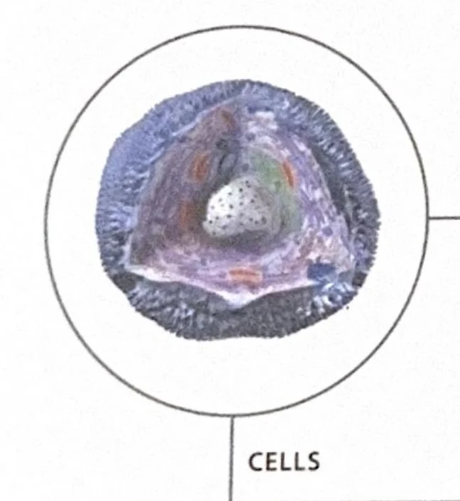
While cells vary wildly in size and shape depending on their specific roles, most share a common fundamental anatomy:
Outer Membrane: A protective barrier that regulates what enters and exits the cell.
Cytoplasm: A jellylike substance that fills the cell and houses internal structures.
Organelles: Specialized "little organs" within the cell that perform specific tasks (e.g., mitochondria for energy production).
Nucleus: A central structure that contains the cell's DNA, acting as the control center.
Diagram of a cell.
Cellular Diversity and Specialization
There are more than 200 different types of cells in the human body. Although every cell contains the same genetic information (with a few exceptions), not all genes are "switched on" in every cell. This pattern of gene expression determines a cell's unique structure and function.
For instance, adipose cells are specialized for fat storage, with much of their interior occupied by a large droplet of semiliquid fat. In contrast, nerve cells (neurons) are electrically excitable, designed to transmit action potentials across long distances to communicate information.
The Red Blood Cell Exception
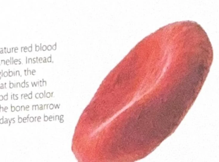
A common "trap" for students is assuming all cells follow the standard nucleus/organelle model. The mature red blood cell is a notable exception. To maximize efficiency in carrying oxygen, these cells are highly specialized: they lack a nucleus and most organelles, instead being packed with hemoglobin, the iron-containing molecule that binds oxygen.
A diagram illustrating a red blood cell, highlighting its biconcave shape and the presence of hemoglobin.
Stem Cells and Differentiation
How does a single fertilized egg become a complex human with specialized brain, muscle, and blood cells? The answer lies in stem cells. Stem cells are unspecialized cells that possess the unique ability to differentiate—meaning they can transform into various specialized tissue cells. This process is influenced by the cell's position in the body and the specific chemical environment it encounters during development.
Tissues: Communities of Cells
When groups of similar cells work together to perform a specific function, they form a tissue. The human body is composed of four primary tissue types:
Epithelial Tissue: These are barrier cells that cover body surfaces and line internal cavities, such as the skin or the lining of the lungs. They can also be specialized to secrete substances (like enzymes) or absorb nutrients.
Connective Tissue: This diverse group provides support and protection for body structures. It includes highly varied forms such as bone, cartilage, fat (adipose), and even blood.
Muscular Tissue: This tissue is specialized for producing movement. It is divided into three main types:
Skeletal Muscle: Attached to bones and responsible for voluntary movements.
Smooth Muscle: Found in the walls of organs like the stomach and blood vessels; it produces involuntary, wave-like contractions.
Cardiac Muscle: Found exclusively in the heart.
Nervous Tissue: This tissue forms the brain, spinal cord, and nerves, facilitating rapid internal communication by transmitting electrical impulses.
Deep Dive: Cardiac Muscle Structure
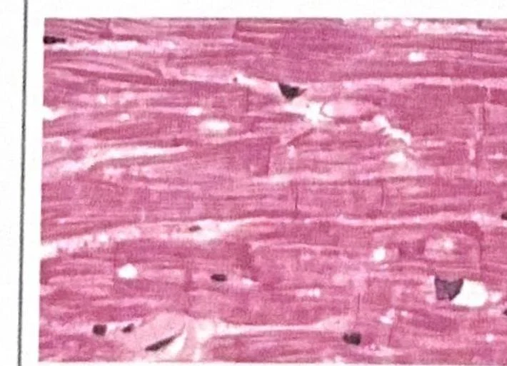
Cardiac muscle is unique in its architecture. Unlike the long, parallel fibers of skeletal muscle, cardiac muscle cells are long, cylindrical, and branched. These branches form junctions with neighboring cells, creating an interconnected network that allows electrical signals to travel rapidly, ensuring the heart's contractions are precisely coordinated.
Microscopic view of cardiac muscle tissue showing striations and nuclei.
Organs: Integrating Tissues
An organ is a discrete structure composed of multiple different tissue types grouped together to perform unique, complex functions. An organ is more than just a collection of cells; it is a functional unit where different tissues cooperate.
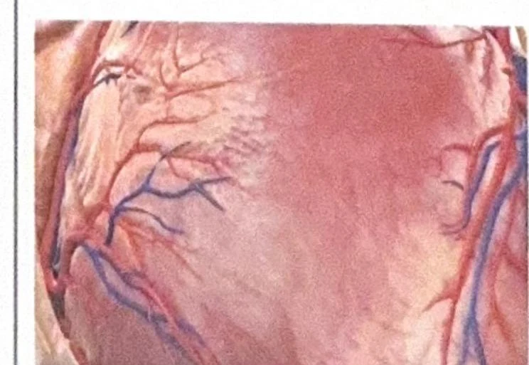
A classic example is the heart:
It uses cardiac muscle tissue to provide the physical pumping action.
It utilizes connective tissue to protect the structure and hold the various parts together.
It employs epithelial tissue to line the internal chambers and cover the valves.
An illustration of the coronary arteries on the surface of a heart.
Body Systems: The Grand Integration
When organs collectively work toward a common purpose, they form a body system. While we often study systems in isolation, they are actually integrated and interdependent. They communicate through complex feedback loops using molecules like hormones and nerve impulses to maintain a state of equilibrium (homeostasis).
The major human body systems include:
System
Primary Function
Key Components
Skeletal
Support and protection
Bones, cartilage, ligaments
Muscular
Movement
Skeletal, smooth, and cardiac muscles
Nervous
Monitoring and control
Brain, spinal cord, nerves
Cardiovascular
Transport of oxygen/nutrients
Heart, blood, blood vessels
Respiratory
Gas exchange (O2 in, CO2 out)
Lungs, trachea, diaphragm
Digestive
Food processing and absorption
Stomach, intestines, liver, pancreas
Urinary
Waste removal and fluid balance
Kidneys, bladder, urethra
Endocrine
Hormonal communication
Glands (e.g., thyroid, pancreas)
Note on Interdependence: Consider the cardiovascular and respiratory systems. The respiratory system brings oxygen into the lungs, but it is the cardiovascular system that actually transports that oxygen to the trillions of cells throughout the body. One cannot function effectively without the other.
By organizing from the microscopic level of the cell to the macroscopic level of the system, the human body creates a seamless transition from simple chemical reactions to the complex, living organism.
Keep reading for free.
Create an account to finish this reading and make it part of your study routine.