Human and Veterinary Tooth and Periodontal Anatomy
An overview of the anatomy of teeth and their supporting structures, including the jaw, articulation, internal tooth components (pulp and dentin), external protective layers (enamel and cementum), and the periodontium (alveolar bone, periodontal ligaments, and gingiva). It also covers directional terms used in dentistr
Understanding the anatomy of the tooth and its supporting structures is fundamental to dental health and veterinary medicine. Whether examining a human or an animal, the oral cavity is a complex system of hard tissues, soft tissues, and specialized joints that work together to facilitate feeding, speech, and structural integrity. This article explores the internal components of the tooth, the protective layers that encase it, and the periodontal structures that anchor the teeth within the jaw.
The Jaw and Articulation
The teeth are housed within the jawbones, which are divided into the upper and lower sections. The maxilla constitutes the upper jaw and is a complex structure made up of many bones. The roof of the mouth is formed by the hard and soft palates. The mandible is the lower jaw.
These two structures are connected and allow for movement through the temporomandibular joint (TMJ). The functionality of this joint is critical for the mechanical processes of biting and chewing.
Internal Tooth Anatomy: Pulp and Dentin
A tooth is not a solid, inert object; it is a living organ with a complex internal environment. At the very center, or the "core" of the tooth, lies the pulp.
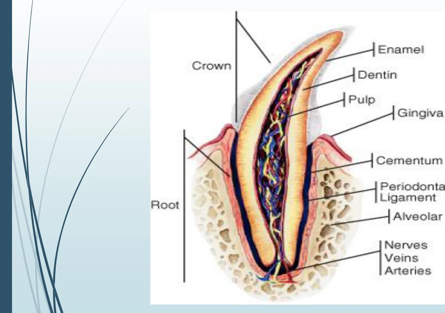
Diagram illustrating the different parts of a tooth, including the crown, root, enamel, dentin, pulp, gingiva, cementum, periodontal ligament, alveolar bone, nerves, veins, and arteries.
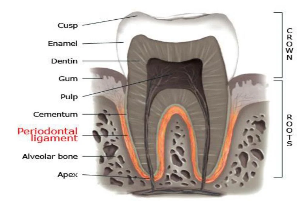
The pulp consists of connective tissue, nerves, and blood vessels that provide nourishment to the tooth. Most of these vital vessels and nerves enter the tooth through the apex, which is the bottom of the root. A unique feature of the pulp is the presence of specialized cells called odontoblasts, which are responsible for forming dentin.
Surrounding the pulp is the dentin, which makes up the bulk of the tooth. While dentin is as hard as bone, it is softer than enamel. It serves a sensory function, allowing the tooth to detect touch, heat, and cold. Dentin is categorized into two types:
Primary dentin: Formed before the tooth erupts.
Secondary dentin: Continually formed throughout the life of the tooth.
As secondary dentin is produced, the pulp chamber gradually reduces in size.
External Protective Layers: Enamel and Cementum
To protect the dentin and pulp, the tooth is encased in specialized hard tissues. The type of tissue covering the dentin depends on whether you are looking at the crown or the root.
Enamel
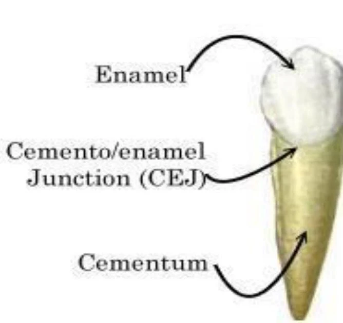
The crown of the tooth is encased in enamel. Enamel is the hardest tissue found in mammals. It is formed before the tooth erupts; however, once eruption occurs, the formation of enamel stops, and the tissue is gradually lost over the lifetime of the tooth. Because of its hardness, it is also susceptible to chipping.
Cementum
The root of the tooth is covered by cementum. This is a hard, calcified tissue that is slowly formed throughout the life of the tooth. Cementum plays a vital role in supporting the tooth within the jaw and assisting in root repair.
Diagram illustrating the enamel, cemento-enamel junction (CEJ), and cementum of a tooth.
The Periodontium: Supporting Structures
The "periodontium" refers to the tissues that surround and support the teeth. This includes the bone, the ligaments, and the gums.
Alveolar Bone and Periodontal Ligaments
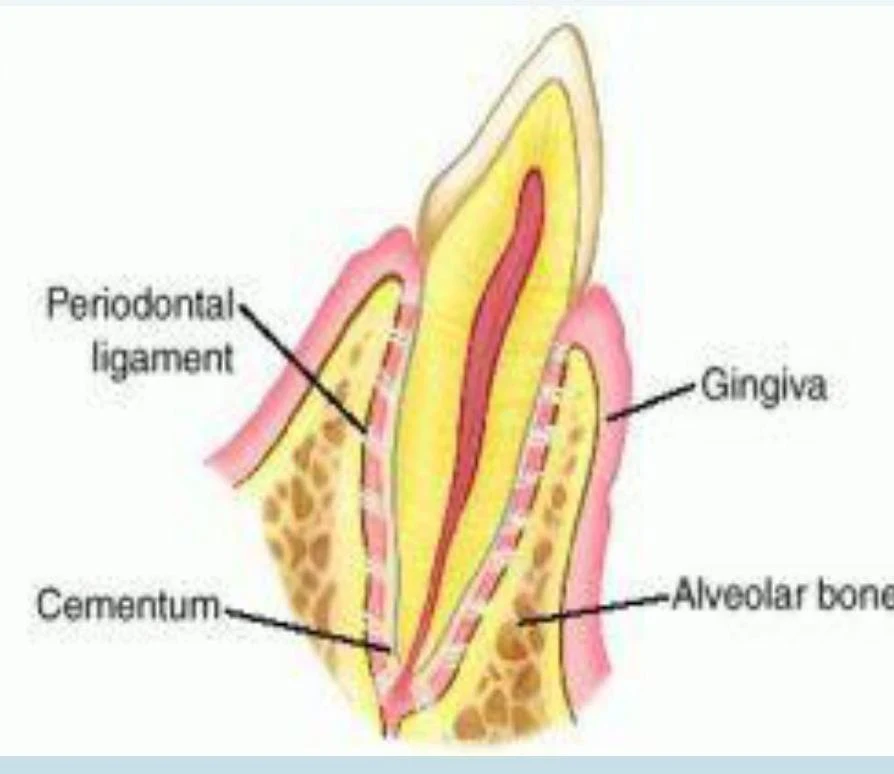
The alveolar bone forms the jaw and creates the sockets into which the roots of the teeth extend.
Diagram illustrating the structure of a tooth, including the periodontal ligament, cementum, gingiva, and alveolar bone.
The tooth is held securely within these sockets by the periodontal ligaments. This tough tissue attaches the cementum of the tooth directly to the alveolar bone.
Diagram illustrating the anatomy of a tooth, including its crown and roots, with labels for various structures such as enamel, dentin, pulp, cementum, periodontal ligament, and alveolar bone.
Gingiva and the Sulcus
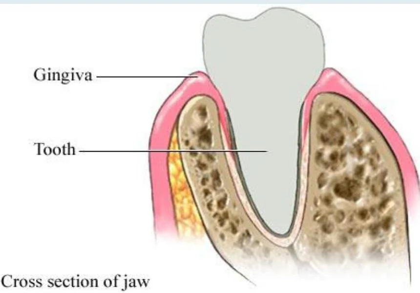
The gingiva, commonly known as the "gums," is the soft tissue that covers the remainder of the periodontium. Where the gingiva is directly attached to the tooth, it is harder and more tightly bound, allowing it to withstand the intense forces of chewing.
The anatomical relationship between the tooth and the gingiva is precise:
Sulcus: The area or space between the free gingiva and the tooth.
Pocket: If the space between the free gingiva and the tooth increases, it is referred to as a pocket.
Cross section of jaw showing tooth and gingiva.
Lateral Canals
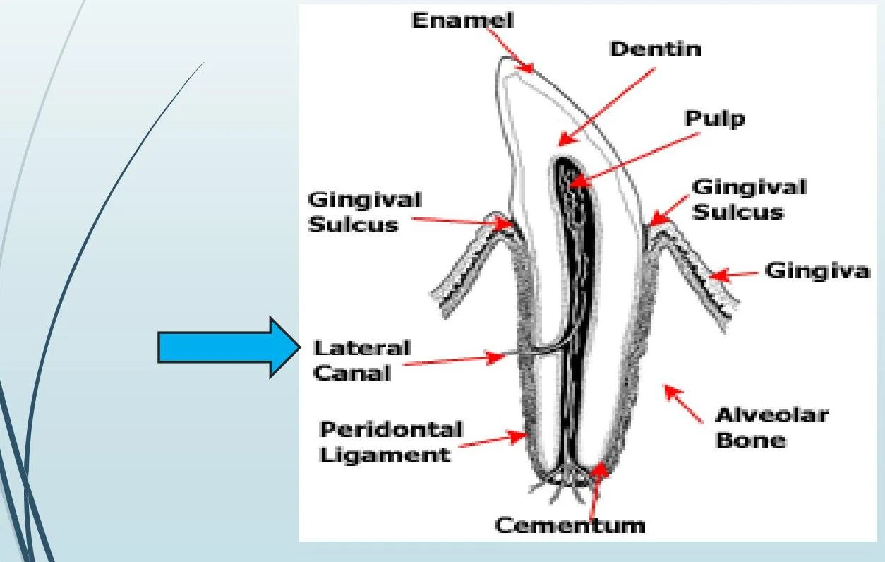
In addition to the main blood vessels entering through the apex, there are lateral canals. These are very small channels that connect the root pulp to the periodontal tissue, providing a pathway for small blood vessels.
Diagram illustrating the anatomy of a tooth, including enamel, dentin, pulp, gingival sulcus, gingiva, lateral canal, periodontal ligament, cementum, and alveolar bone.
Directional Terms in Dentistry
To accurately describe dental health, clinicians use specific directional terms to locate surfaces and positions relative to the tooth and gums.
Key terms include:
Coronal: Toward the crown of the tooth.
Apical: Toward the apex (the tip of the root).
Supragingival: Above the gum line.
Subgingival: Below the gum line.
Study Tip: When performing oral charting or taking radiographs, always use these directional terms to ensure precise communication regarding the location of decay, fractures, or periodontal disease.
Keep reading for free.
Create an account to finish this reading and make it part of your study routine.