Human Anatomy: The Skeletal System and Connective Tissue Histology
Explore the human skeletal system, from its macroscopic divisions (axial and appendicular) to its microscopic composition of specialized connective tissues like cartilage and bone. Learn about the extracellular matrix, collagen fibers, and the classification of joints (fibrous, cartilaginous, and synovial).
The human skeletal system is much more than a rigid framework of bones; it is a complex, dynamic organ system composed of specialized tissues, intricate microstructures, and diverse joint classifications. Understanding the skeletal system requires a dual approach: looking at the macroscopic organization of the body and the microscopic histology of the tissues that provide strength, flexibility, and movement.
Connective Tissue Composition and the Extracellular Matrix
At the microscopic level, the skeletal system is built upon connective tissues. Unlike epithelial tissue, which consists of tightly packed cells, connective tissue is characterized by a significant amount of extracellular matrix (ECM) that surrounds resident cells.
The composition of the extracellular matrix is critical to the function of the tissue. It generally consists of two main components: ground substance and protein fibers.
This illustration depicts the components of connective tissue, including the extracellular matrix with protein fibers (elastic, collagen, reticular) and ground substance, as well as resident cells like mesenchymal cells, macrophages, adipocytes, and fibroblasts, alongside a blood vessel.
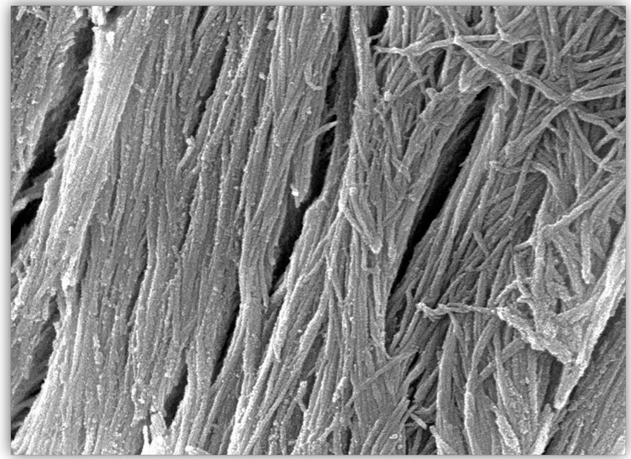
The protein fibers, most notably collagen fibers, provide the structural integrity and tensile strength necessary for bones and cartilage to withstand mechanical stress.
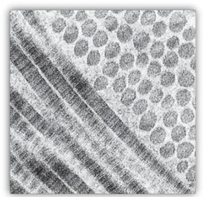
Micrograph showing collagen fibrils and proteoglycans.
Scanning electron micrograph of carbon nanotubes.
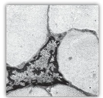
A transmission electron micrograph shows the nucleus of a cell surrounded by cytoplasm.
Study Tip: When examining histology slides, look for the orientation of collagen fibers. In dense regular connective tissue, these fibers are often arranged in parallel bundles to resist tension in a single direction.
Cartilage and Bone Microstructure
The skeletal system utilizes different types of specialized connective tissues to meet varying functional demands, such as shock absorption and structural support.
Cartilage Types
Cartilage is a resilient tissue that lacks blood vessels (avascular). Two primary types are essential to skeletal function:
Hyaline Cartilage: This is the most common type of cartilage. It features chondrocytes (cartilage cells) located within small spaces called lacunae, embedded in a smooth, glassy-looking matrix.
Fibrocartilage: Designed for high-stress areas, fibrocartilage contains a much denser matrix of collagen fibers, providing significantly more strength than hyaline cartilage.
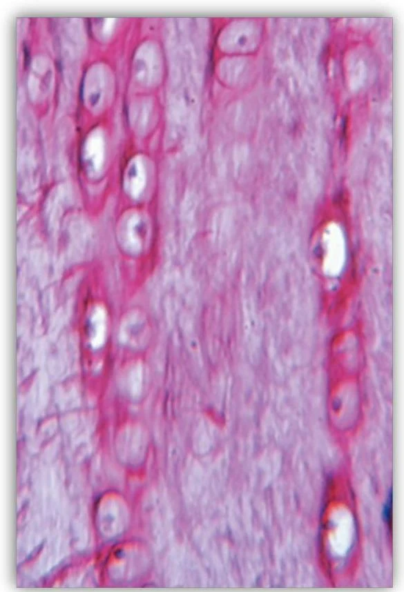
Microscopic view of chondrocytes within lacunae in cartilage tissue.
Bone Tissue
Bone is a highly specialized connective tissue with a unique microstructure. Unlike cartilage, bone is characterized by a fibrous collagen matrix that is mineralized to provide hardness.
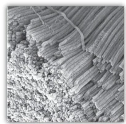
Scanning electron micrograph showing collagen fibers.
Macroscopic Anatomy: Axial and Appendicular Skeletons
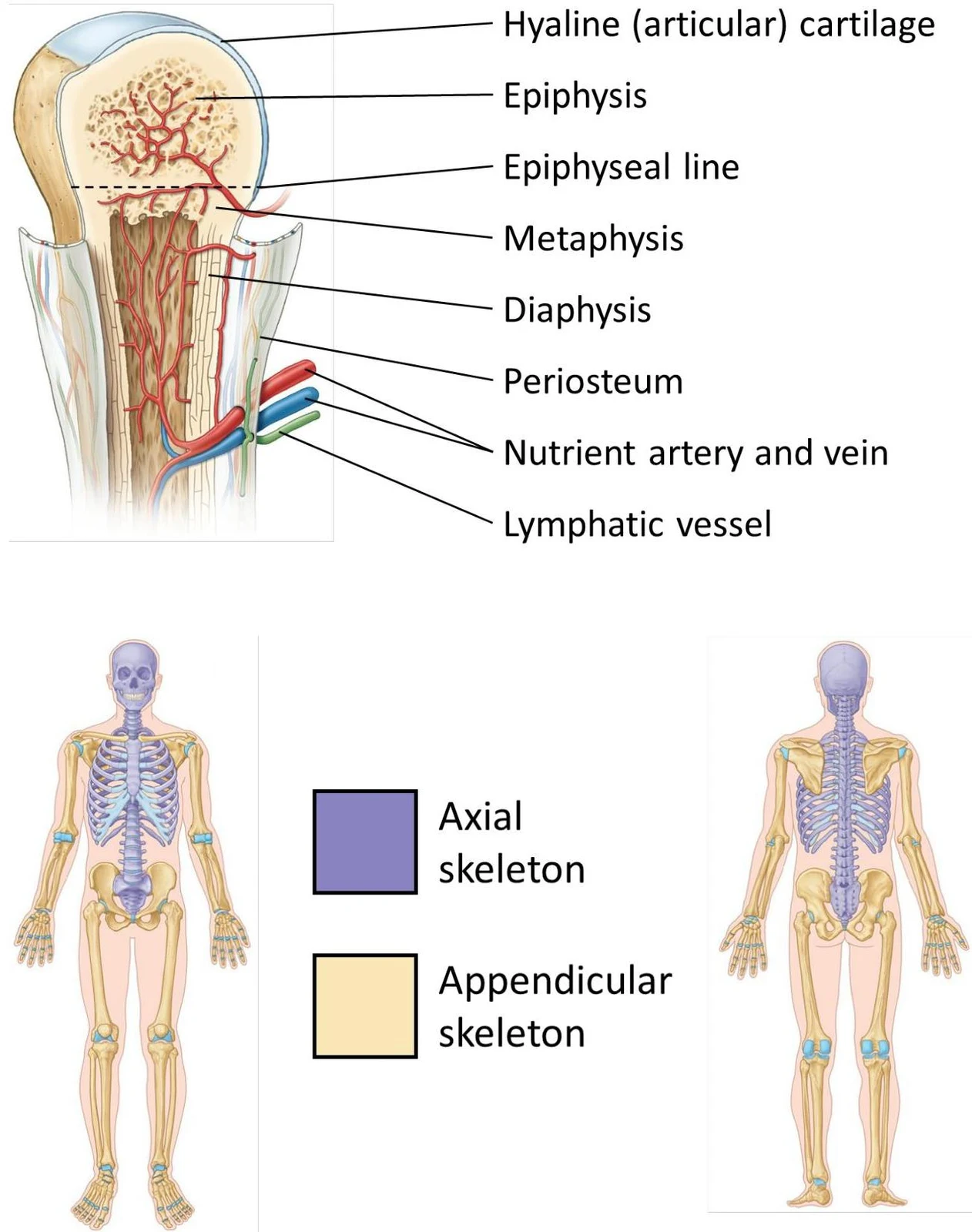
On a larger scale, the human skeleton is divided into two main functional divisions: the axial skeleton and the appendicular skeleton.
Axial Skeleton: Forms the central axis of the body, including the skull, vertebral column, and thoracic cage.
Appendicular Skeleton: Consists of the bones of the upper and lower limbs, as well as the girdles (pectoral and pelvic) that attach the limbs to the axial skeleton.
Diagrams illustrating the anatomy of a long bone and the axial and appendicular skeletons.
Classification of Joints
Joints (articulations) are the points where two or more bones meet. They are classified based on their structure (the material binding them) and their function (the degree of movement allowed).
Fibrous Joints
Fibrous joints are joined by dense connective tissue and allow for little to no movement. Examples include:
Suture: Found only between the bones of the skull.
Syndesmosis: A joint where bones are connected by a ligament or fibrous membrane.
Cartilaginous Joints
These joints are held together by cartilage and are categorized into two types:
Primary Cartilaginous Joint (Synchondrosis): These involve a bar or plate of hyaline cartilage connecting the bones. An example is the epiphyseal plate in a growing long bone.
Secondary Cartilaginous Joint (Symphysis): These involve a fibrocartilage pad between bones, such as the intervertebral discs in the spine.
Synovial Joints
Synovial joints are the most common and mobile joints in the body. They are characterized by the presence of a fluid-filled space that reduces friction. Key components include:
Articular Cartilage: Covers the bone surfaces to reduce friction.
Joint Cavity: The space containing synovial fluid.
Joint Capsule: An envelope surrounding the joint.
Synovial Membrane: The inner lining of the capsule that secretes synovial fluid.
There are six distinct types of synovial joints, each providing different mechanical advantages, such as hinge or ball-and-socket movements.
Keep reading for free.
Create an account to finish this reading and make it part of your study routine.