An overview of the human circulatory system, including the different types of blood vessels, the two main circulatory circuits (pulmonary and systemic), and the vascularization of key body regions like the brain, digestive tract, and lower extremities. It also touches upon clinical aspects like intracranial hemorrhage
The human circulatory system is a sophisticated, pressure-regulated, closed system designed to transport life-sustaining substances throughout the body. It functions as a continuous loop, delivering oxygen, nutrients (such as glucose, amino acids, and vitamins), and hormones to tissues while simultaneously removing metabolic waste products like carbon dioxide, urea, and bilirubin. To achieve this, the body utilizes two distinct circuits that pass through the heart, ensuring that oxygen-rich and oxygen-poor blood remain separate.
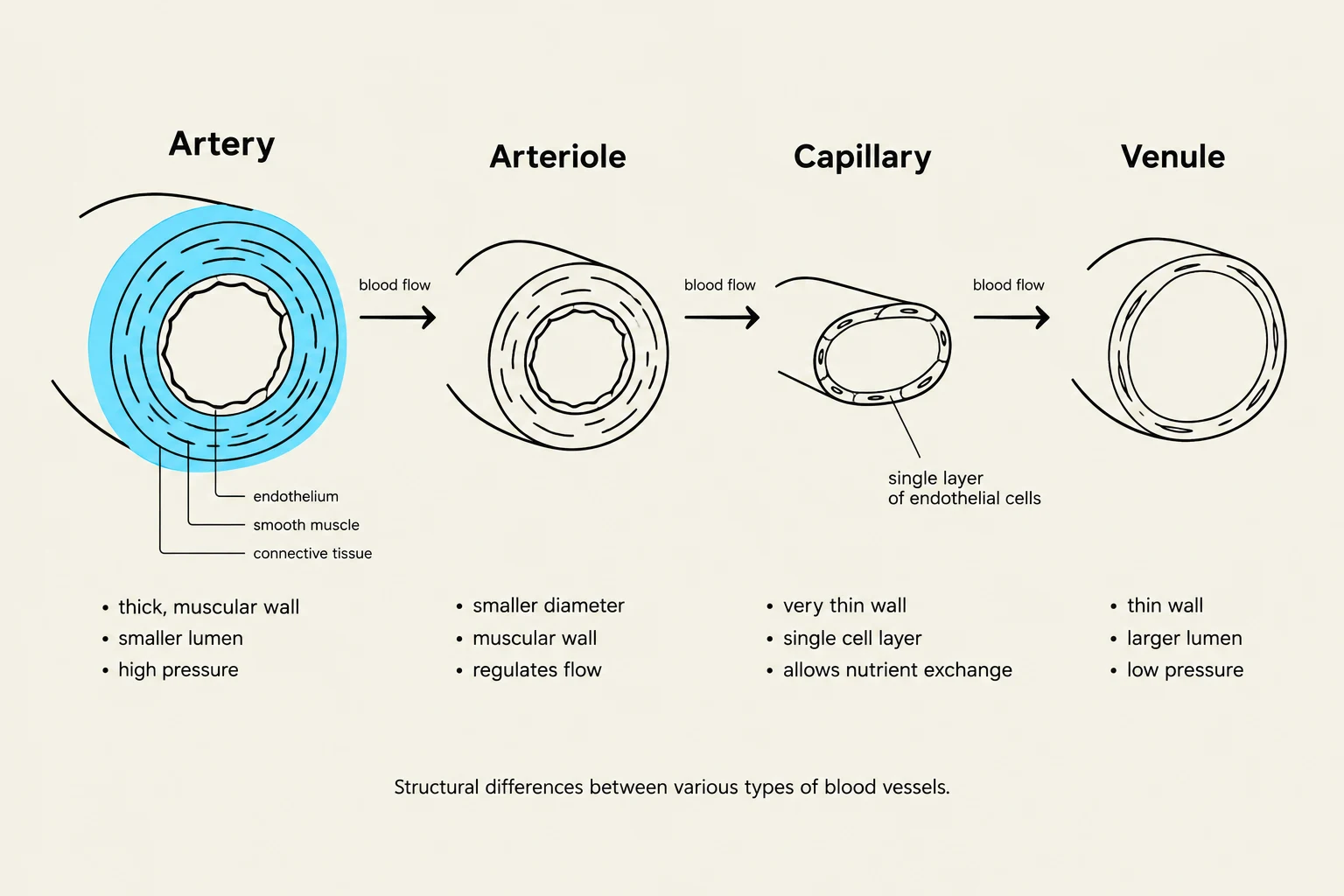
Structural differences between arteries, arterioles, capillaries, and venules, illustrating their varying wall thickness, lumen size, and pressure.
Classes of Blood Vessels
Blood vessels are not uniform; they are specialized structures categorized by their function, wall thickness, and the pressure they must withstand.
Arteries and Arterioles
Arteries are high-pressure vessels that carry blood away from the heart. They possess thick, muscular walls to accommodate the forceful ejection of blood from the ventricles. As arteries branch deeper into tissues, they become arterioles, which are the smallest arterial branches. Arterioles play a critical role in regulating blood pressure and local tissue perfusion through vasoconstriction and vasodilation.
Capillaries and Venules
The ultimate destination of arterial blood is the capillaries. These are the smallest blood vessels and serve as the primary site of exchange between the blood and the interstitial fluid. In the capillaries, oxygen and nutrients move into the tissues, while waste products move into the bloodstream.
Once exchange is complete, blood enters venules, which collect blood from the capillary beds. Venules then merge into larger veins, which are low-pressure vessels that return blood to the heart. Unlike arteries, veins have thinner walls and often contain valves to prevent the backflow of blood.
Circulatory Circuits
The human body operates via two primary circuits:
Pulmonary Circulation: This circuit pumps deoxygenated blood from the right side of the heart to the lungs to pick up oxygen and release carbon dioxide.
Systemic Circulation: This circuit pumps oxygenated blood from the left side of the heart to the rest of the body, including the brain, organs, and extremities.
Cerebral Circulation and Hemorrhage
The brain requires a constant, redundant supply of blood to maintain function. It is vascularized via the brachiocephalic trunk, common carotid arteries, and vertebral arteries.
The Circle of Willis
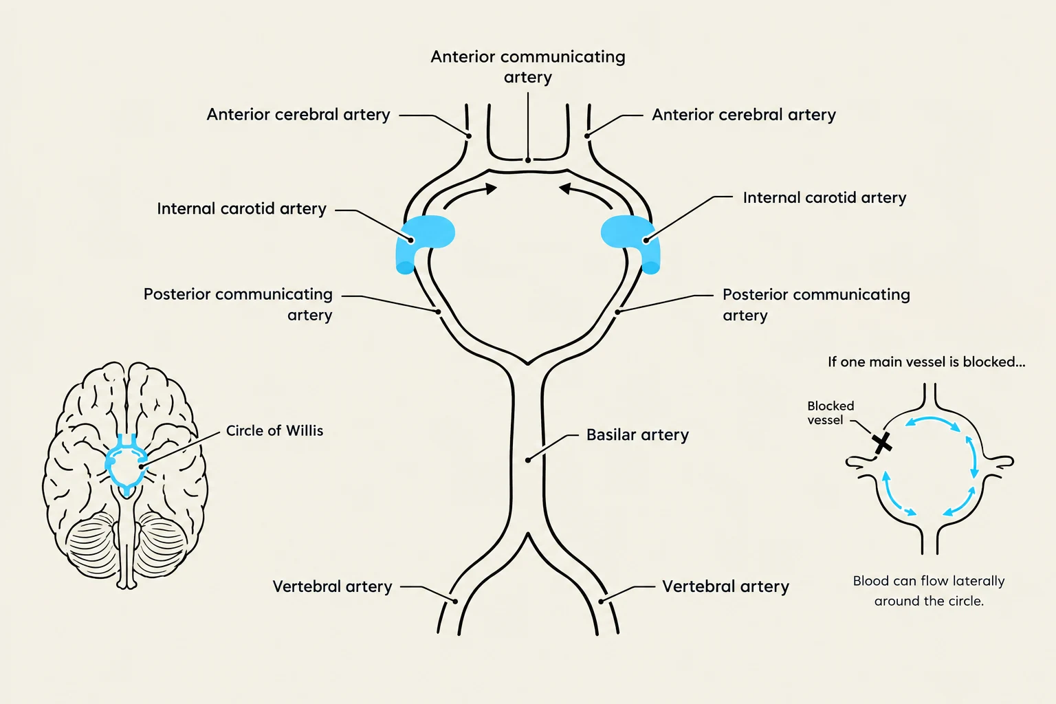
Diagram illustrating the Circle of Willis, a crucial arterial network at the base of the brain, showing blood flow pathways and the effect of a blocked vessel.
A vital anatomical feature of the brain is the Circle of Willis (the cerebral arterial circle). This is an anastomosis—a connection between vessels—that interconnects the internal carotid arteries and the basilar artery. This circular arrangement ensures that if one vessel becomes blocked, blood can be rerouted from other vessels to prevent brain tissue death.
Venous Drainage and Dural Sinuses
Venous blood leaves the brain through superficial cerebral veins and smaller veins that empty into a network of dural sinuses. Key sinuses include the superior and inferior sagittal sinuses, the transverse sinus, and the straight sinus. This blood eventually drains into the jugular veins.
Clinical Distinctions in Intracranial Hemorrhage
Understanding the anatomical layers of the brain is essential for diagnosing types of bleeding:
Type of Hemorrhage
Anatomical Location
Common Cause/Vessel Involved
Epidural
Between the skull and the dura mater
Middle meningeal artery
Subdural
Between the dura and the arachnoid mater
Ruptured bridging veins
Subarachnoid
Between the arachnoid and the pia mater
Aneurysmata
Intracerebral
Within the brain tissue itself
Various
Exam Tip: If a patient presents with a skull fracture and rapid loss of consciousness, suspect an epidural hemorrhage involving the middle meningeal artery. If the patient shows gradual disorientation without trauma, consider a subdural hemorrhage caused by bridging veins.
Systemic and Visceral Circulation
The descending aorta is the main artery supplying the lower half of the body, and it is divided into the thoracic aorta and the abdominal aorta.
Vascularization of the Digestive Tract
The abdominal aorta provides blood to the digestive organs through three major branches:
Celiac Trunk: Supplies the stomach, spleen, pancreas, and duodenum.
Superior Mesenteric Artery: Supplies the small intestine, the ascending colon, and the proximal two-thirds of the transverse colon.
Inferior Mesenteric Artery: Supplies the distal one-third of the transverse colon, the descending colon, and the rectum.
The Portal System
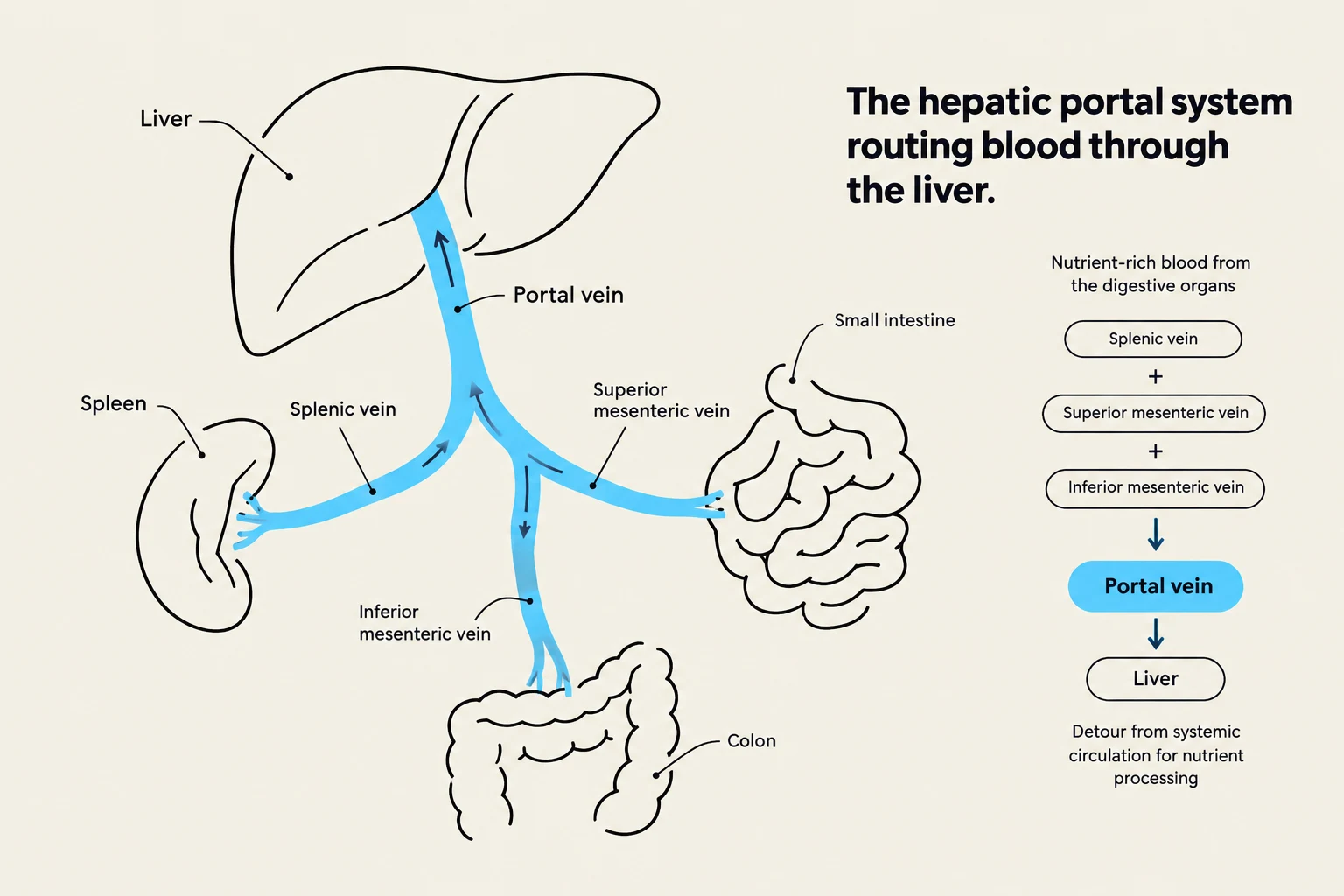
Diagram illustrating the hepatic portal system, which routes nutrient-rich blood from the digestive organs and spleen to the liver for processing.
A unique aspect of abdominal anatomy is the portal system. Instead of returning directly to the heart, blood from the intestines is routed through the portal vein to the liver. The portal vein is formed by the convergence of the splenic, superior mesenteric, and inferior mesenteric veins. This allows the liver to "clear" or process nutrients and toxins from the blood before it enters general circulation.
In cases of liver disease (such as chronic alcoholism), blood flow can be obstructed, leading to portocaval anastomoses—alternative pathways between the portal and systemic systems. Clinical congestion in these areas can result in visible signs like the "medusa sign".
Lower Extremity Blood Flow
Blood travels to the lower limbs through a specific sequence of arterial branching:
Common Iliac Artery
Internal and External Iliac Arteries
Femoral Artery
Deep Femoral Artery
Popliteal Artery
Anterior and Posterior Tibial Arteries
Fibular Artery
Clinical Note: To prevent blood clots (thrombi) from traveling to vital organs, or to assist venous return against gravity, it is important to maintain muscle activity. This is why "muscle pumping" in the legs is essential for circulation.
Keep reading for free.
Create an account to finish this reading and make it part of your study routine.