Host Defenses and the Lymphatic System: An Overview
An overview of the human immune system's three lines of defense, including innate barriers, internal defenses, and adaptive immunity. It also details the structure and function of the lymphatic system and its role in immune surveillance.
The human body is constantly under siege by a diverse array of pathogens, including bacteria, viruses, and fungi. To survive, we rely on an interconnected network of cells, fluids, and organs designed for a singular purpose: immunosurveillance. This process involves searching for, recognizing, and destroying foreign material to maintain health. This complex defense mechanism is organized into three distinct lines of defense, ranging from general physical barriers to highly specific, memory-based responses.
Diagram illustrating the three lines of host defense: physical and chemical barriers, innate immune response, and adaptive immune response.
The Three Lines of Defense
The immune system does not act as a single unit but rather as a layered defense strategy. These layers often overlap, providing redundant protection to ensure that if one fails, another is ready to take over.
1. The First Line: Innate/Nonspecific Barriers
The first line of defense consists of physical and chemical barriers that attempt to block the entry of microbes at their portals of entry. This line is considered "innate" because it is inborn and does not require prior exposure to a pathogen to function.
Physical/Anatomical Barriers: The skin (specifically the keratinized stratum corneum) acts as an impervious, waterproof shield. Mucous membranes in the respiratory, digestive, and urinary tracts trap microbes, while mechanisms like the "mucociliary escalator" in the lungs move trapped particles toward the pharynx to be expelled.
Chemical Barriers: These include substances that inhibit or kill microbes, such as lysozyme in tears and saliva, the acidic pH of stomach acid (HCl), and antimicrobial peptides like defensins.
The Microbiome: Our normal microbiota serves as a structural and chemical barrier by competing for nutrients and altering local pH to prevent pathogen colonization.
2. The Second Line: Innate/Nonspecific Internal Defenses
If a pathogen breaches the first line, the second line of defense is activated. These are internal processes that kick in quickly to contain the threat. Key components include:
Phagocytosis: The process where "eating cells" (phagocytes) like neutrophils and macrophages ingest and destroy microbes.
Inflammation: A localized response characterized by four cardinal signs: Rubor (redness), Calor (warmth), Tumor (swelling), and Dolor (pain).
Fever: A systemic increase in body temperature triggered by pyrogens, which can inhibit microbe growth and speed up immune reactions.
Antimicrobial Proteins: This includes the complement system (a cascade of proteins that can lyse bacteria) and interferons (proteins that help limit viral multiplication).
3. The Third Line: Adaptive/Specific Immunity
The third line is the "acquired" response. Unlike the innate system, it is highly specific to particular antigens—unique molecular markers (proteins or sugars) displayed by cells or viruses. This line relies on specialized lymphocytes: B cells, which provide humoral immunity through antibodies, and T cells, which manage cell-mediated immunity. The hallmark of the third line is its ability to provide long-term immunity through memory.
Exam Tip: PAMPs vs. PRRs
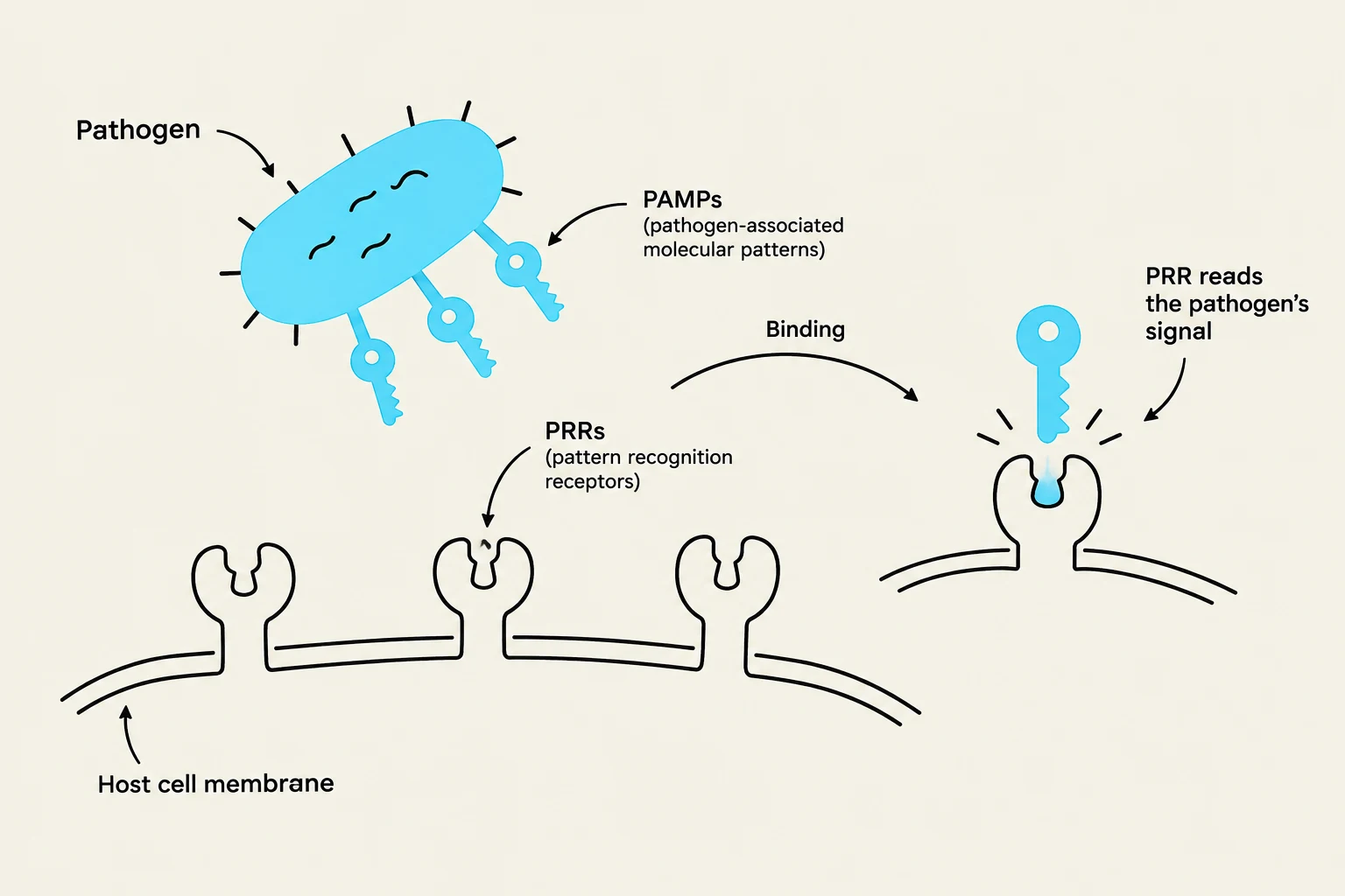
To distinguish between these on a test, remember: P-A-MP is the "Pathogen's Ad"—the microbe advertises itself with Pathogen-Associated Molecular Patterns. P-R-R is the "Receptor Reads"—the host's Pattern Recognition Receptors read those signals.
Immune Recognition: Self vs. Nonself
This diagram illustrates how pathogen-associated molecular patterns (PAMPs) on a pathogen bind to pattern recognition receptors (PRRs) on a host cell membrane, signaling the presence of the pathogen.
How does the body know what to attack? The innate immune system uses Pattern Recognition Receptors (PRRs), such as Toll-like receptors (TLRs), to detect Pathogen-Associated Molecular Patterns (PAMPs). PAMPs are molecular "red flags" found on microbes but not in mammals.
When the system correctly identifies "self" antigens, it ignores them. However, when "nonself" antigens are detected, the immune system marks them for destruction. If the system mistakenly attacks "self" proteins, it results in an autoimmune disorder, such as rheumatoid arthritis.
The Lymphatic System
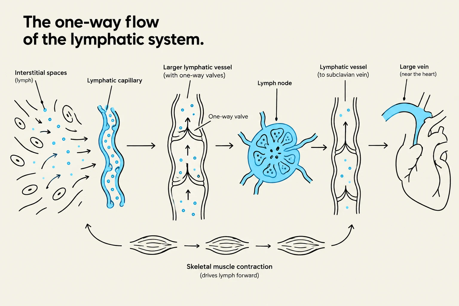
Diagram illustrating the one-way flow of the lymphatic system, showing interstitial spaces, lymphatic capillaries, larger lymphatic vessels with valves, lymph nodes, lymphatic vessels leading to the subclavian vein, and a large vein near the heart. The diagram also indicates that skeletal muscle contraction drives lymph forward.
The lymphatic system is an interconnected network of vessels, cells, and organs that permeates every tissue. It serves three primary functions: returning extracellular fluid (ECF) to the blood, providing a route for immune surveillance, and acting as a "drain-off" for inflammation.
Lymph and Vessels
Lymph is a plasma-like fluid that carries white blood cells (especially lymphocytes), fats, and cellular debris, but notably contains no red blood cells.
Unlike the circulatory system, which has a central pump (the heart), the lymphatic system is a one-way system. Lymph flows from the extremities toward the heart, driven only by skeletal muscle contraction.
Clinical Connection: Because lymph movement relies on muscle contraction, patients who are immobilized (like those in hospital beds) are at risk for swelling in the hands and feet. This is why compression sleeves are often used to assist fluid return.
Lymphoid Organs
Immune organs are divided into two categories based on their role in the life cycle of a lymphocyte:
Primary Lymphoid Organs (Sites of Birth and Maturation)
Red Bone Marrow: The site of all blood cell production (hematopoiesis) and the specific site where B cells undergo maturation.
Thymus: The specialized site where T cells undergo maturation and develop specificity.
Secondary Lymphoid Organs (Sites of Activation)
This is "where the action happens"—where immune cells encounter pathogens and activate.
Lymph Nodes: Small, bean-shaped structures that filter lymph.
Spleen: A large organ in the upper left abdomen that filters blood, removing worn-out red blood cells and pathogens.
Associated Lymphoid Tissues (ALT/MALT/GALT): These are bundles of lymphocytes located under skin and mucosal surfaces to provide "first-strike" protection. Examples include the tonsils and Peyer's patches (located in the ileum of the small intestine).
Summary Table: Key Immune Components
Component
Type
Primary Function
Neutrophils
Innate
Most common WBC; primary phagocyte in acute infection
Macrophages
Innate
Large phagocytes; differentiate from monocytes
B Cells
Adaptive
Mature in Bone marrow; produce antibodies
T Cells
Adaptive
Mature in Thymus; cell-mediated immunity
Complement
Innate
Protein cascade that causes cell lysis via MAC
Keep reading for free.
Create an account to finish this reading and make it part of your study routine.