Histology: The Study of Tissues and Embryonic Development
Explore the tissue level of biological organization, the four primary tissue types (epithelial, connective, muscle, nervous), and their origins from embryonic germ layers. Learn about early human embryonic development and body membranes.
To understand how the human body functions, we must look beyond individual cells and examine how they work together. This is the essence of histology—the microscopic study of tissues. A tissue is defined as a group of similar cells that share a common embryonic origin and a common function, all bound together by an intercellular substance. In the hierarchy of biological organization, tissues represent the level above the cellular level and below the organ level.
Early Human Embryonic Development
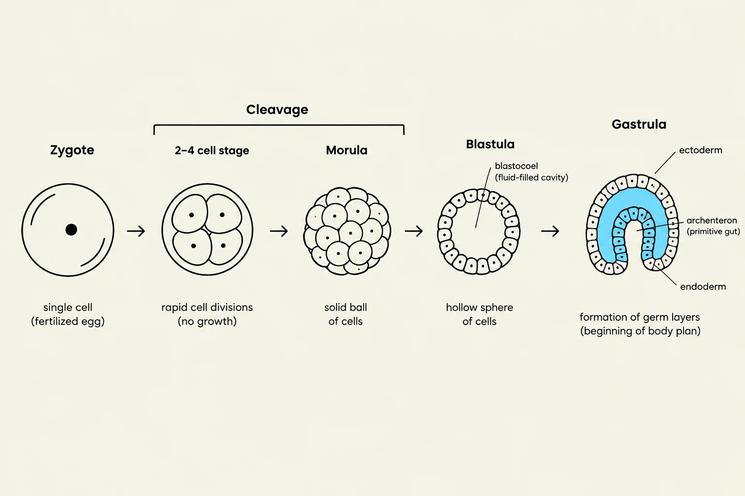
The journey from a single cell to a complex organism begins with fertilization, which creates a zygote. The first eight weeks post-fertilization is known as the embryonic period, a critical window where the basic body plan emerges and organs begin to form.
During this period, the embryo undergoes several key processes:
Cleavage: Rapid mitotic cell divisions that increase cell number without increasing the overall size of the embryo.
Morula: A solid ball of cells resulting from cleavage.
Blastula: A hollow ball of cells that forms after the morula stage.
Gastrulation: A transformative process where the embryo reorganizes into three distinct primary germ layers.
Morphogenesis: The process by which cell migration and mitosis change the physical shape of the embryo.
Differentiation: The process by which cells acquire specific structures and specialized functions.
Diagram illustrating the stages of early embryonic development, including zygote, cleavage, morula, blastula, and gastrula, showing the formation of germ layers.
The Primary Germ Layers and Their Derivatives
The three germ layers formed during gastrulation serve as the "blueprints" for all adult tissues:
Ectoderm: Gives rise to the nervous system and the epidermis (skin), including hair and skin glands.
Mesoderm: Forms the "middle" structures, including muscle tissues (trunk and limbs), the heart, blood, bones, ligaments, kidneys, gonads, and the walls of the digestive and respiratory tracts. It also forms the visceral and parietal serosa.
Endoderm: Forms the inner linings, specifically the lining of the gut and respiratory systems, as well as the glands associated with them.
Exam Tip: While most tissues have a specific germ layer origin, remember that epithelium is a descriptive term. Epithelial tissue can actually be derived from all three germ layers. This is a common "trick" question on exams!
The Four Primary Tissue Types
Once differentiation is complete, the body is composed of four fundamental tissue types: epithelial, connective, muscle, and nervous.
1. Epithelial Tissue
Epithelial tissues cover body surfaces, line internal cavities, and form glands. They are characterized by being avascular (lacking blood vessels), meaning they must rely on diffusion from underlying tissues for nutrients. They are attached to a basement membrane, which serves as a foundation and a site for stem cells to facilitate healing and growth.
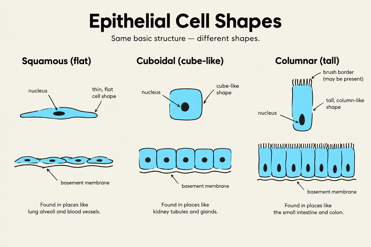
Epithelia are classified by cell shape and the number of layers:
Simple: A single layer, ideal for diffusion (e.g., simple squamous in the lungs) or transport (e.g., simple columnar in the intestines).
Stratified: Multiple layers, providing protection against abrasion (e.g., stratified squamous in the skin or mouth).
Specialized types: Includes pseudostratified (often ciliated, found in the trachea) and transitional (capable of stretching, found in the bladder).
Glandular Epithelium is a specialized form that secretes substances. Endocrine glands release hormones directly into the blood, while exocrine glands release substances through ducts onto local surfaces.
Intercellular Junctions
Because epithelial cells must act as barriers, they are held together by specialized junctions:
Tight Junctions: Create a seal to prevent leakage, common in transport epithelia like the intestines.
Desmosomes: Provide strong mechanical attachment, essential in the epidermis and cardiac muscle.
Gap Junctions: Allow ions and small molecules to pass between cells, facilitating communication in cardiac and smooth muscle.
Hemidesmosomes: Connect cells to the underlying basement membrane.
Diagram illustrating the three main shapes of epithelial cells: squamous (flat), cuboidal (cube-like), and columnar (tall), along with their locations in the body.
2. Connective Tissue
Connective tissue is the most diverse group. Unlike epithelium, its cells are usually widely separated by an abundant extracellular matrix (ECM) consisting of ground substance and fibers.
The Extracellular Matrix Components:
Ground Substance: Can vary from liquid (blood) to gel or solid (bone).
Fibers: Includes collagen (tough and pliable), elastin (stretchy), and reticular (fine framework).
Major Categories:
Loose Connective Tissue: Includes areolar (a general packing tissue) and adipose (fat storage for energy and insulation).
Dense Connective Tissue: Includes dense regular (parallel collagen in tendons) and dense irregular (interwoven collagen in the dermis).
Specialized Tissues: Includes cartilage (slow-healing due to lack of blood supply), bone (solid matrix), and blood (liquid matrix/plasma).
3. Muscle Tissue
Muscle tissue is specialized for contraction to produce movement. All muscle types utilize the proteins actin and myosin to facilitate shortening.
Skeletal Muscle: Long, cylindrical, multinucleated cells that are striated (striped) and under voluntary control.
Cardiac Muscle: Branched, striated cells found in the heart. They are involuntary and connected by intercalated discs containing gap junctions.
Smooth Muscle: Spindle-shaped, non-striated cells found in the walls of hollow organs (like the GI tract). They are involuntary and produce wave-like contractions called peristalsis.
4. Nervous Tissue
Nervous tissue enables rapid communication throughout the body. It consists of two main cell types:
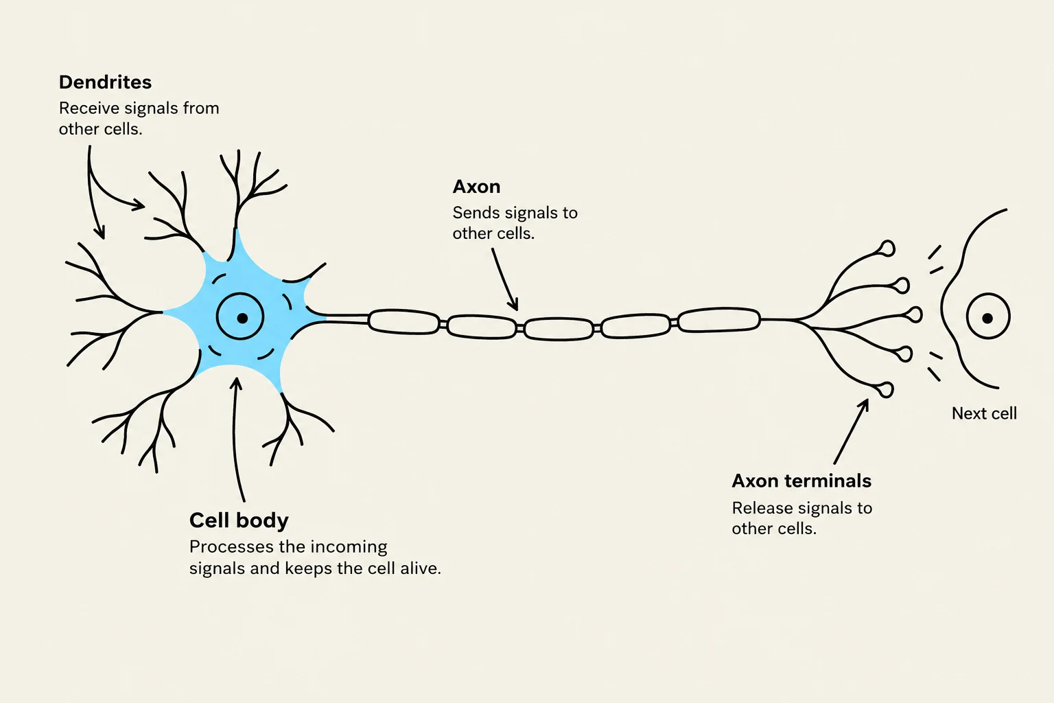
Neurons: The functional units that conduct electrical signals via processes called dendrites (receive signals) and axons (send signals).
Neuroglia: Supporting cells that protect and nourish the neurons.
Diagram illustrating a neuron, including its dendrites, cell body, axon, and axon terminals, and its connection to a next cell.
Body Membranes and Cavities
Membranes are organs composed of an epithelial layer sitting atop a layer of connective tissue.
Mucous Membranes (Mucosa): Line cavities open to the exterior (e.g., mouth, digestive tract, respiratory tract).
Serous Membranes: Line closed internal cavities. They consist of a parietal layer (lining the cavity wall) and a visceral layer (covering the organ). They secrete serous fluid to reduce friction. Examples include the pleura (lungs), pericardium (heart), and peritoneum (abdominal cavity).
Synovial Membranes: Line joint capsules and produce lubricating synovial fluid.
Cutaneous Membrane: The skin, which covers the external surface of the body.
Understanding these tissues and how they originate from the embryonic germ layers provides the foundation for studying all human organ systems.
Keep reading for free.
Create an account to finish this reading and make it part of your study routine.