An in-depth exploration of the histological structures of the oral cavity and teeth, covering their development, composition, and the specialized cells involved in their formation and maintenance.
The oral cavity serves as the gateway to the digestive system, where food is ingested, masticated, and lubricated by saliva before swallowing. To withstand the mechanical rigors of chewing, the oral cavity is lined with specialized stratified squamous epithelium, which can be keratinized (such as on the hard palate and gingiva) or nonkeratinized (such as on the cheeks and floor of the mouth). Within this environment, the teeth stand out as highly specialized, mineralized structures designed for mechanical breakdown. Understanding their histology requires an exploration of the complex interplay between ectodermal and mesenchymal cell lineages during embryological development.
Dental Anatomy and Dentition
An adult human possesses a permanent dentition consisting of 32 teeth. These are arranged in two bilaterally symmetric arches: the maxillary (upper) and mandibular (lower) arches. Before these permanent teeth erupt, they are preceded by a set of primary teeth, also known as deciduous or "milk" teeth, which are eventually shed.
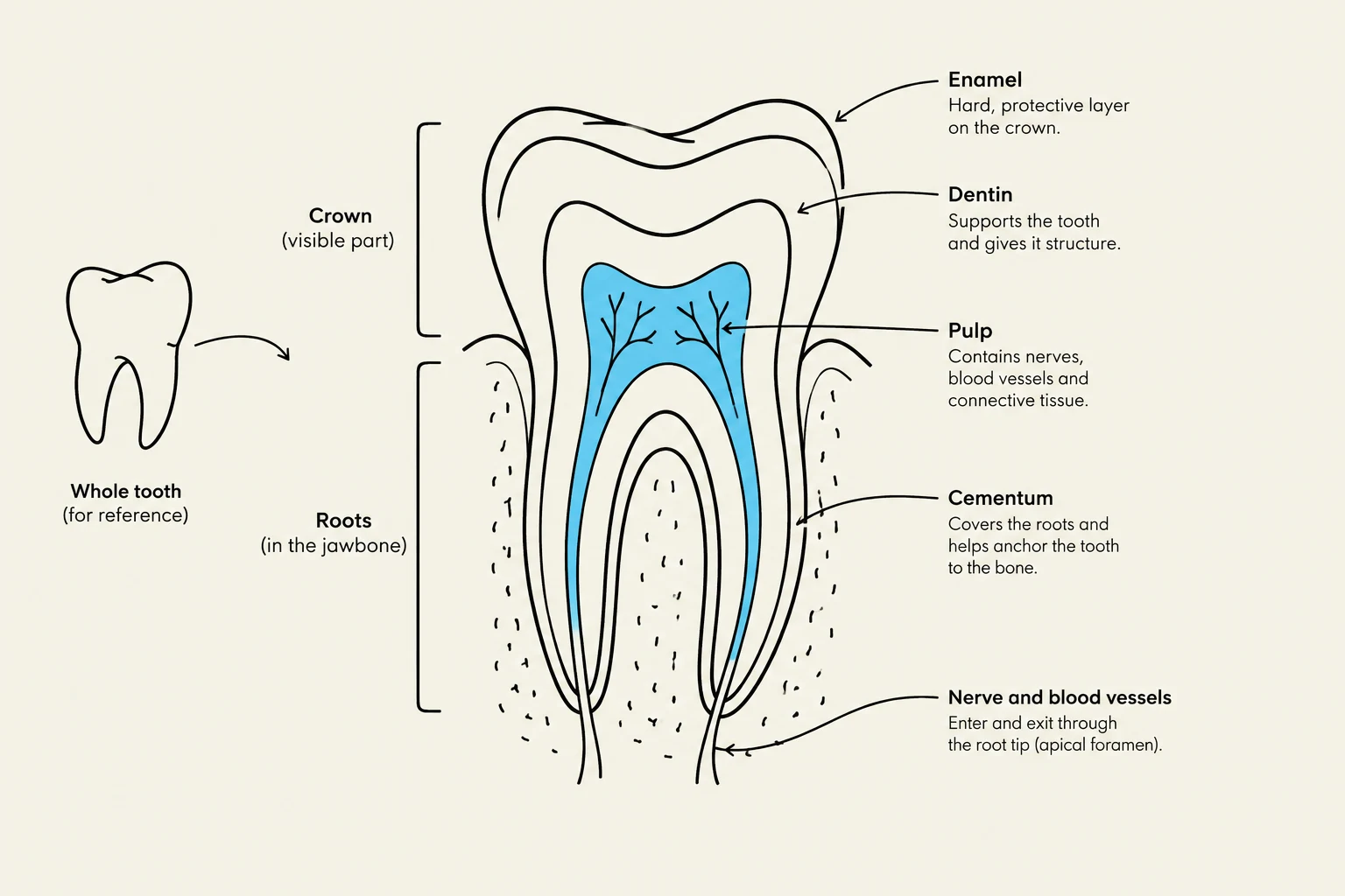
While the number of teeth varies between primary and permanent sets, the fundamental histological structure remains consistent across all teeth. A tooth is generally divided into three regions:
The Crown: The visible portion covered by enamel.
The Neck: The constricted area where the enamel and cementum meet at the gingiva.
The Root: The portion anchored within the alveolar bone, covered by cementum.
Exam Tip: Remember that while enamel is the hardest substance in the body, it is non-vital (contains no cells) once fully formed, whereas the dentin and pulp remain biologically active throughout life.
Diagram illustrating the different parts of a tooth, including the enamel, dentin, pulp, cementum, and nerve and blood vessels.
Embryological Development: The Enamel Organ and Dental Papilla
Tooth formation is a masterpiece of cellular induction. It begins in the embryo when the ectodermal epithelium of the oral cavity invaginates into the underlying mesenchyme. This invagination forms the enamel organ, a specialized epithelial structure shaped like a wine glass.
The development relies on two primary cell populations:
Ameloblasts: Derived from the ectoderm, these tall, polarized cells form the innermost layer of the enamel organ.
Odontoblasts: Derived from neural crest cells located within the mesenchymal dental papilla (the tissue inside the concave portion of the enamel organ).
The relationship between these cells is interdependent: as the odontoblasts begin to secrete the initial organic matrix, their presence induces the ameloblasts to begin secreting enamel.
Enamel: The Hardest Substance
Enamel is the most highly mineralized tissue in the human body, composed of approximately 96% calcium hydroxyapatite. Notably, enamel contains very little protein and lacks collagen, which distinguishes it from bone and dentin. Because it lacks a cellular component, enamel cannot repair itself once damaged.
The structural unit of enamel is the enamel rod (or prism). These are interlocking columns, approximately 5 micrometers in diameter, that extend through the entire thickness of the enamel layer. The growth of these rods is guided by amelogenin, a small structural protein secreted by the ameloblasts via their ameloblast (Tomes) process.
Clinical Correlation: The addition of fluoride to toothpaste helps incorporate fluoride into the hydroxyapatite crystals, forming fluoroapatite. This makes the enamel more resistant to the acidic dissolution caused by microorganisms.
Dentin and the Role of Odontoblasts
Dentin is a calcified tissue that is harder than bone but less mineralized than enamel, consisting of roughly 70% hydroxyapatite. Unlike enamel, dentin is an organic-rich tissue containing Type I collagen and glycosaminoglycans (GAGs).
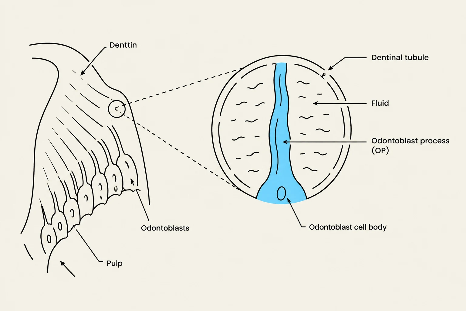
The production of dentin is driven by odontoblasts, which are tall, polarized cells that line the pulp cavity. The process follows a specific sequence:
Predentin Secretion: Odontoblasts secrete an unmineralized organic matrix called predentin.
Mineralization: Within approximately one day, hydroxyapatite crystals form within the predentin, transforming it into hard dentin.
Tubule Formation: As the dentin layer thickens, the long apical extensions of the odontoblasts (odontoblast processes) remain trapped within microscopic channels called dentinal tubules.
Diagram illustrating the structure of dentin, pulp, odontoblasts, and dentinal tubules within a tooth.
These dentinal tubules are critical for tooth sensitivity. Unmyelinated nerve fibers from the pulp extend into these tubules, allowing the tooth to respond to stimuli like heat, cold, or acid as pain.
The Pulp Cavity and Periodontium
The center of the tooth contains the pulp cavity, which is filled with pulp: a well-vascularized and highly innervated mesenchymal connective tissue. The pulp serves as the life-support system for the tooth, providing nutrients through blood vessels and sensory information through nerves that enter the tooth via the apical foramina at the root tips.
While enamel formation ceases shortly before the tooth erupts, odontoblasts persist throughout adult life, continuing to produce dentin and maintaining the matrix.
To keep the tooth anchored in the jaw, the periodontium provides a complex support system:
Cementum: An avascular, bone-like layer that covers the dentin of the root.
Periodontal Ligament (PDL): A highly cellular, fibrous connective tissue containing bundles of collagen fibers that bind the cementum to the alveolar bone.
Alveolar Bone: The bony socket that houses the tooth, which undergoes continuous remodeling.
Gingiva: The specialized keratinized oral mucosa that surrounds the neck of the tooth.
Keep reading for free.
Create an account to finish this reading and make it part of your study routine.