


The human skeletal system is a complex architecture of living cells embedded within a non-living extracellular matrix (ECM). This matrix provides the necessary strength and support for the body through a delicate balance of organic proteins—primarily collagen—and inorganic mineral salts. When the genetic instructions for synthesizing these components are flawed, the structural integrity of the entire body can be compromised. This article explores two significant hereditary connective tissue disorders: Osteogenesis Imperfecta and Marfan Syndrome, detailing how molecular mutations manifest as profound clinical changes in the skeleton and other organ systems.
Osteogenesis Imperfecta: Brittle Bone Disease
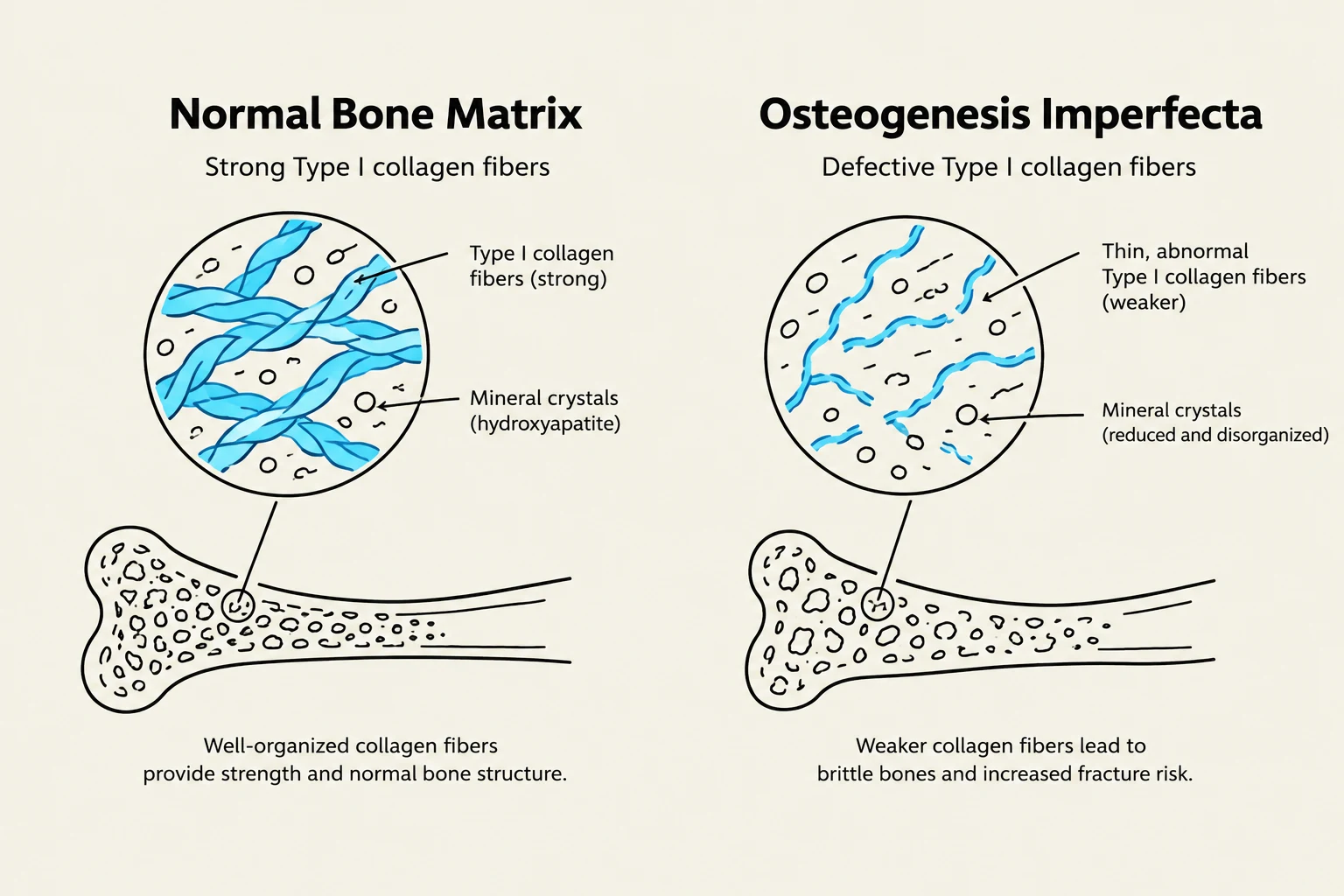
Osteogenesis Imperfecta (OI), often referred to as "brittle bone disease," is a group of hereditary disorders characterized by an abnormal synthesis of Type I collagen. Collagen is the primary organic component of the bone matrix, providing the tensile strength required to prevent bones from shattering under stress.
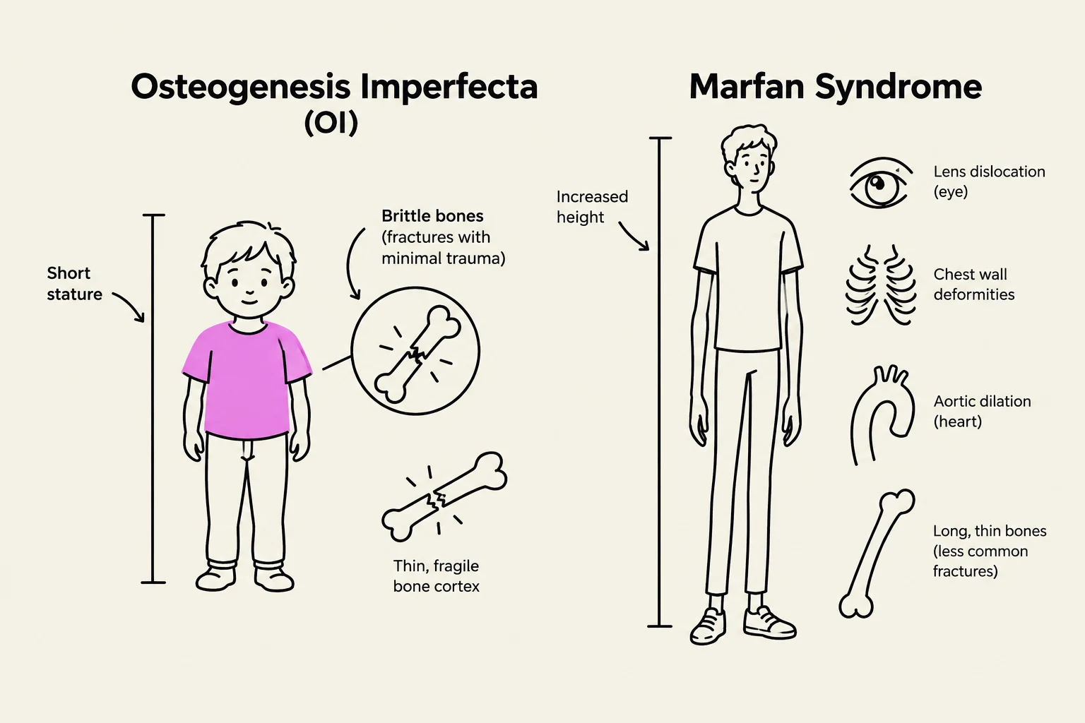
In patients with OI, the defect in collagen production leads to a fundamental morphological change known as osteopenia—a reduction in bone mass and density. Because the bones lack the necessary organic "scaffolding," they become extremely fragile, making them highly susceptible to frequent fractures and dislocations.
Clinical Manifestations and Diagnosis
Beyond skeletal fragility, OI can present with several systemic features depending on the severity of the mutation:
- Blue sclera: A characteristic bluish tint to the whites of the eyes.
- Auditory issues: Deafness resulting from bone abnormalities in the middle ear.
- Dental and growth issues: Abnormal tooth development and short stature.
Diagnosis typically involves a multi-modal approach. Clinicians utilize imaging diagnosis, such as radiography or ultrasound (even in utero), to visualize bone structure and fractures. For definitive confirmation, genetic testing is used to identify specific mutations, and analysis of collagen production by fibroblasts can be performed.
Keep reading for free.
Create an account to finish this reading and make it part of your study routine.
Continue reading freeAlready have an account? Log in
Updated