Hepatobiliary Pathologies and Gallbladder Disorders
An overview of the hepatobiliary system, focusing on congenital abnormalities, gallstones (cholelithiasis), gallbladder inflammation (cholecystitis), and gallbladder cancer. It also covers clinical management, including cholecystectomy.
The hepatobiliary system, comprising the liver and the gallbladder, plays a vital role in digestion, detoxification, and metabolic homeostasis. While the liver performs hundreds of functions, the gallbladder serves as a specialized reservoir that stores and concentrates bile produced by the liver. When the delicate balance of bile composition is disrupted, or when structural abnormalities exist, a variety of pathologies can arise, ranging from manageable stones to life-threatening malignancies and congenital obstructions.
Congenital Abnormalities and Biliary Atresia
Some patients are born with structural variations in their gallbladder anatomy. These congenital abnormalities may include the complete absence of the gallbladder, the presence of a duplicated gallbladder, or an ectopic location where the gallbladder sits outside its normal anatomical position.
A particularly severe congenital condition is biliary atresia. This is an idiopathic, progressive fibrosis of the bile ducts that results in a complete or partial blockage of bile flow. Biliary atresia is a critical clinical concern because it is the most common cause of neonatal jaundice requiring surgical intervention and stands as a leading cause of pediatric liver transplants.
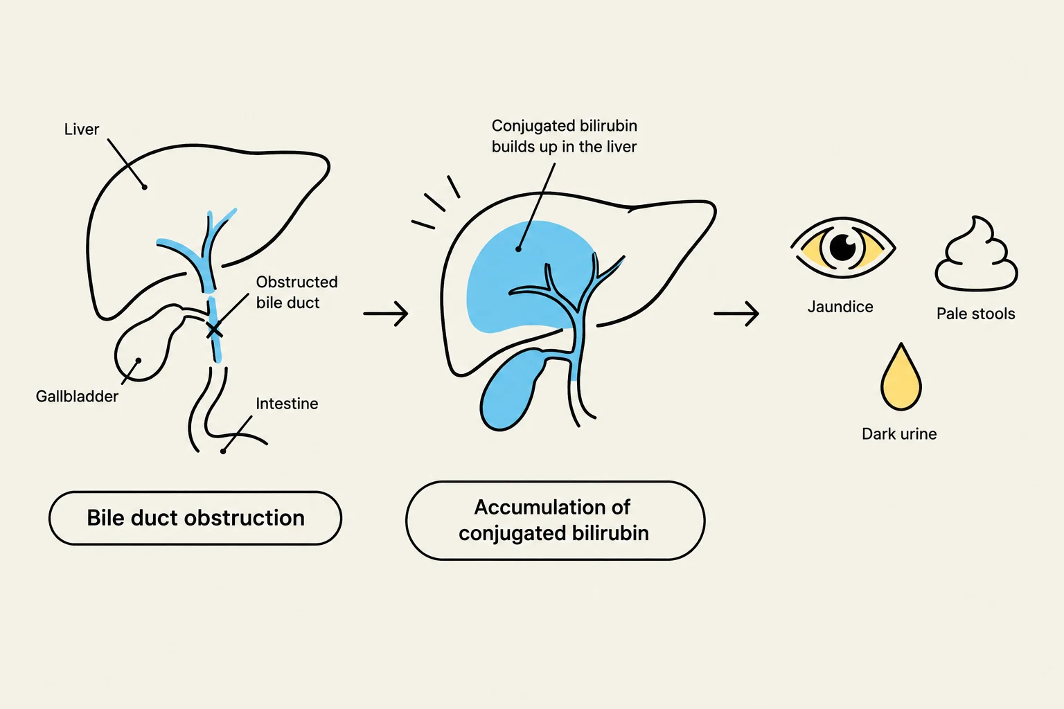
The pathophysiology of biliary atresia follows a predictable, devastating path: the obstruction prevents conjugated bilirubin from being excreted into the GI tract, leading to jaundice, dark urine, and pale stools. If the obstruction is not addressed, the resulting inflammation leads to biliary cirrhosis and, eventually, total liver failure.
Diagram illustrating bile duct obstruction, leading to the accumulation of conjugated bilirubin in the liver, and resulting in symptoms like jaundice, pale stools, and dark urine.
Cholelithiasis: The Pathophysiology of Gallstones
Cholelithiasis, or the presence of gallstones, affects approximately 10% of the adult population. The development of these stones is driven by abnormalities in bile composition and cholestasis (the impairment of bile flow), which causes the precipitation of cholesterol and bilirubin.
Gallstones are categorized by their chemical composition:
Cholesterol stones: The most prevalent type, accounting for roughly 80% of cases, occurring when bile becomes supersaturated with cholesterol.
Black pigment stones: These are associated with hemolysis (the breakdown of red blood cells) and increased hemoglobin turnover.
Brown pigment stones: These are often linked to bacterial or parasite colonization within the biliary tract and are more common in developing countries.
Risk factors for developing stones include obesity, female gender, genetics (particularly in Indigenous populations), the use of oral contraceptives, and malabsorption disorders.
Clinical Note: Biliary Colic
While many patients with gallstones remain asymptomatic, others experience biliary colic. This is characterized by intense, episodic pain in the epigastric region or the Right Upper Quadrant (RUQ), often accompanied by nausea and vomiting.
Cholecystitis: Inflammation of the Gallbladder
When gallstones cause irritation or obstruction, the gallbladder becomes inflamed, a condition known as cholecystitis. There are three primary clinical presentations:
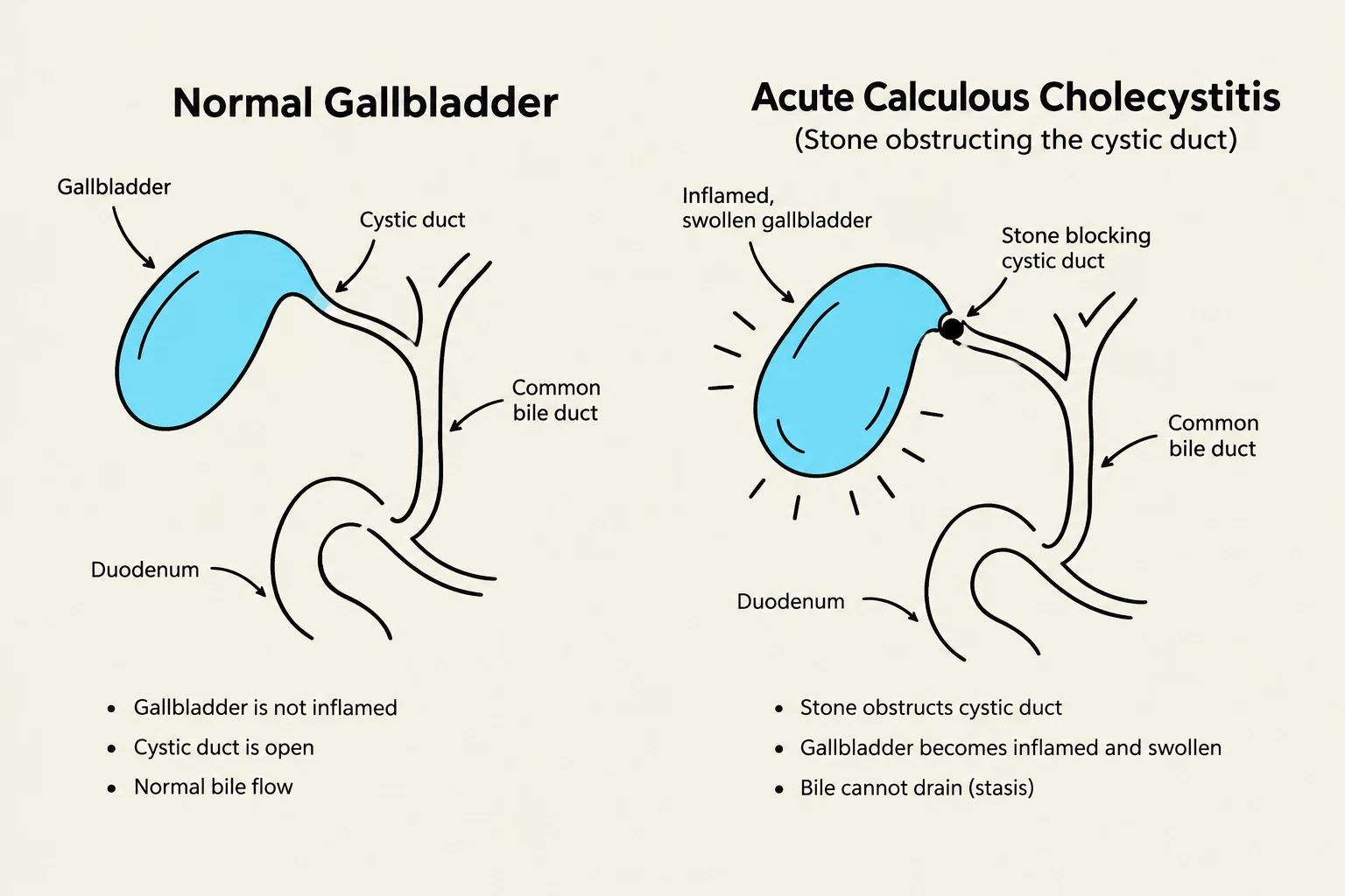
Acute Calculous Cholecystitis: This accounts for 90% of acute cases. It occurs when a gallstone obstructs the cystic duct, causing concentrated bile to irritate the gallbladder wall chemically. This triggers an inflammatory response that can lead to ischemia (restricted blood flow) and distension.
Acute Acalculous Cholecystitis: Representing about 10% of acute cases, this occurs in patients without gallstones. It is typically seen in critically ill patients—such as those suffering from major trauma, severe burns, or those receiving total parenteral nutrition.
Chronic Cholecystitis: This results from long-term mechanical irritation by stones or recurrent bouts of acute inflammation, eventually leading to gallbladder fibrosis.
If left untreated, cholecystitis can progress to severe complications, including gallbladder perforation, sepsis, peritonitis, or abscess formation.
Diagrams illustrating a normal gallbladder and a gallbladder affected by acute calculous cholecystitis due to a stone obstructing the cystic duct.
Gallbladder Cancer
Gallbladder cancer is an uncommon but highly fatal malignancy. One of its most significant clinical features is its strong correlation with pre-existing gallbladder disease; approximately 80% of patients with gallbladder cancer also have cholelithiasis.
Several key risk factors increase the likelihood of developing this cancer:
Porcelain gallbladder: This refers to the calcification of the gallbladder wall.
Gallbladder polyps: Growths on the inner lining of the gallbladder.
The prognosis for gallbladder cancer remains poor, with a 5-year survival rate of only approximately 10%.
Clinical Management: Cholecystectomy
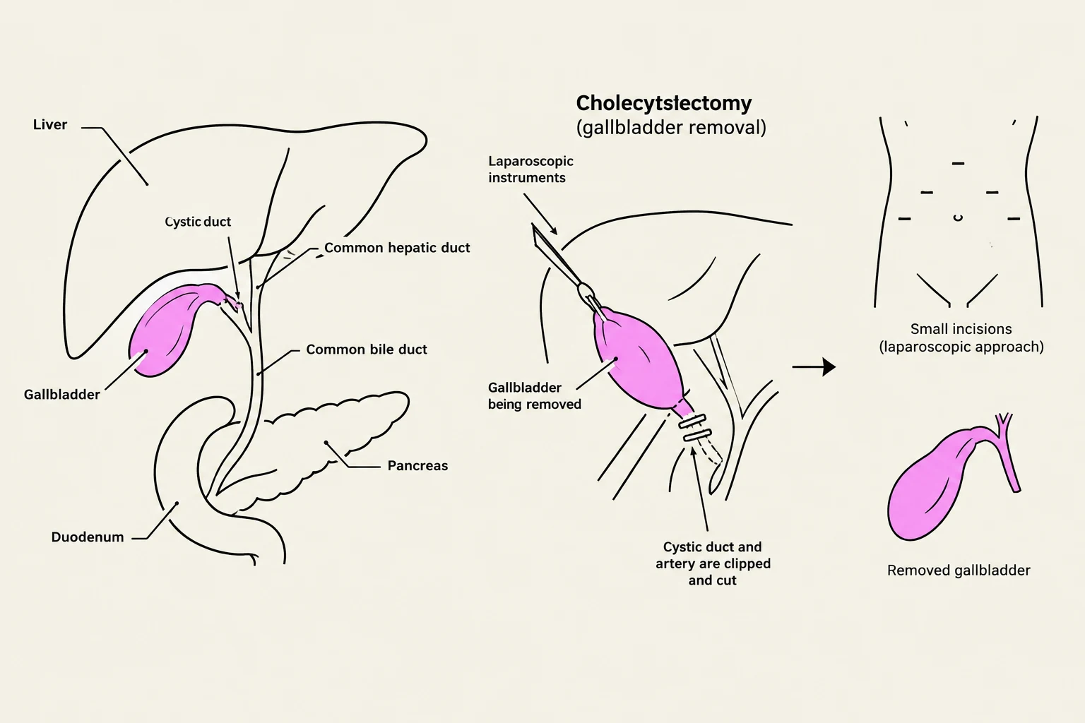
For patients suffering from symptomatic cholelithiasis or cholecystitis, the standard surgical treatment is a cholecystectomy—the surgical removal of the gallbladder. This can be performed via traditional open surgery or, more commonly, through laparoscopic techniques.
Exam Tip: Differential Diagnosis
When evaluating a patient with RUQ pain, fever, and leukocytosis, always consider the distinction between calculous and acalculous cholecystitis. The presence or absence of stones on an ultrasound is the primary diagnostic differentiator.
Diagram illustrating the anatomy of the gallbladder and surrounding organs, and the steps involved in a laparoscopic cholecystectomy (gallbladder removal).
Keep reading for free.
Create an account to finish this reading and make it part of your study routine.