


Hemoglobin is a complex protein essential for oxygen transport in the blood. Its production is a highly regulated process that requires the precise coordination of iron availability, heme biosynthesis, and the synthesis of globin polypeptide chains. Because hemoglobin is the primary vehicle for oxygen delivery, any disruption in its synthesis or structural integrity can lead to significant clinical pathologies, such as anemia or hemoglobinopathies.
The Components of Hemoglobin
To understand how hemoglobin is built, we must first look at its three fundamental components: protoporphyrin IX, iron, and globin.
- Protoporphyrin IX: A complex organic ring structure.
- Iron: Specifically in the ferrous () state, which is capable of binding oxygen.
- Globin: The protein portion consisting of polypeptide chains.

Heme Biosynthesis: A Cellular Journey
Heme synthesis is a multi-step enzymatic process that occurs within nucleated red blood cell precursors (normoblasts). It is important to note that mature red blood cells lack mitochondria and therefore can no longer synthesize hemoglobin.
The Role of Mitochondria and Cytosol
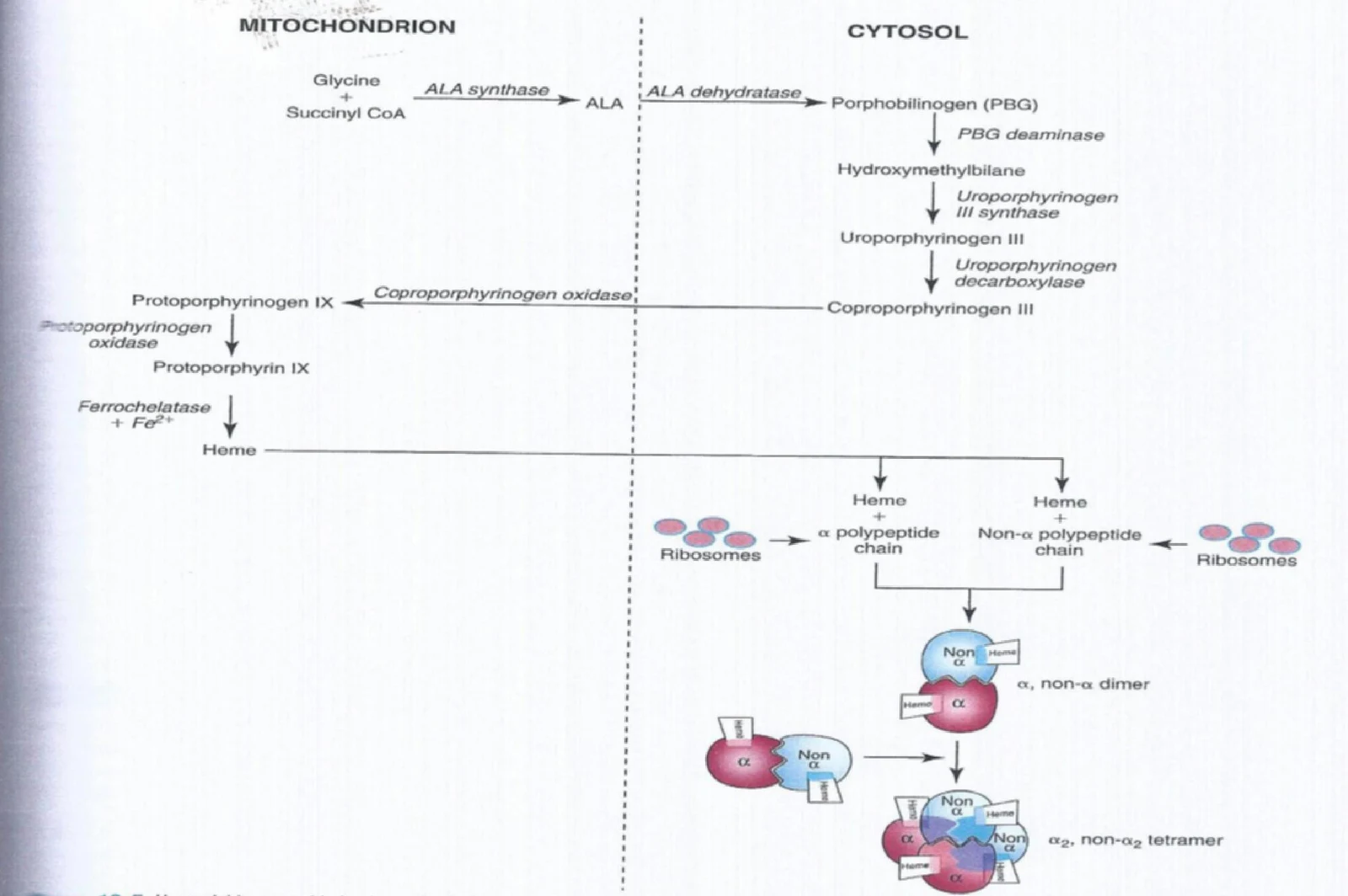
The synthesis pathway is unique because it shuttles between two cellular compartments: the mitochondrion and the cytosol.
- In the Mitochondrion: The process begins here with the assembly of glycine and succinyl CoA. This step is catalyzed by the enzyme aminolevulinic acid (ALA) synthase to form ALA.
- In the Cytosol: The ALA undergoes several enzymatic transformations to eventually form coproporphyrinogen, which is then converted into protoporphyrinogen IX.
- Return to the Mitochondrion: The final stages occur back in the mitochondria. The enzyme ferrochelatase catalyzes the insertion of ferrous iron () into the protoporphyrin ring to create the final heme molecule.
Keep reading for free.
Create an account to finish this reading and make it part of your study routine.
Continue reading freeAlready have an account? Log in
Updated