Hemoglobin Synthesis and Destruction: A Biological Overview
An in-depth explanation of hemoglobin synthesis, its essential components like iron and protoporphyrin, and the processes of hemoglobin destruction through extravascular and intravascular hemolysis. It also covers iron salvage and recycling mechanisms.
Hemoglobin is the essential protein within red blood cells (RBCs) responsible for oxygen transport. The life cycle of hemoglobin is a finely tuned metabolic process involving complex synthesis in the bone marrow and a highly regulated destruction phase once the RBCs reach senescence. Understanding the transition from healthy hemoglobin production to the various pathways of hemolysis—both intravascular and extravascular—is critical for diagnosing hematologic disorders such as anemia, jaundice, and hemoglobinopathies.
Hemoglobin Synthesis
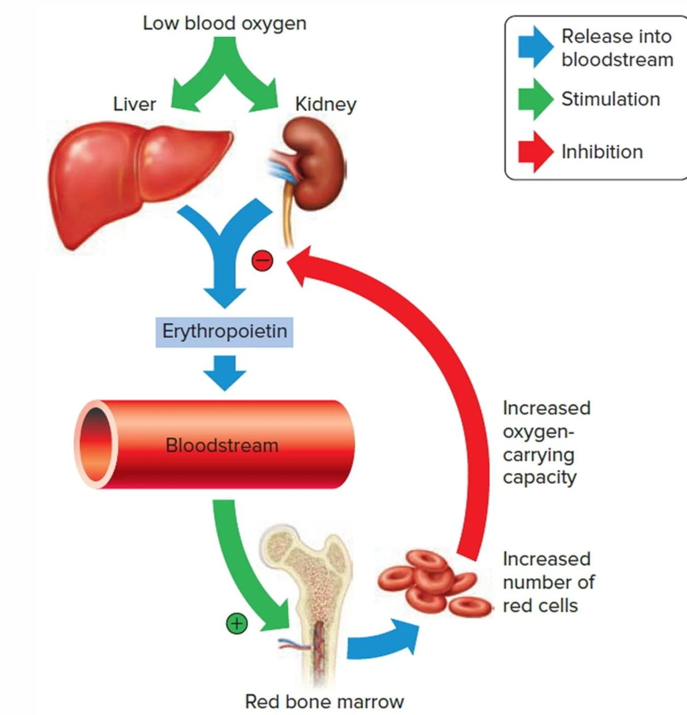
Hemoglobin synthesis is primarily stimulated by tissue hypoxia. When oxygen levels in the tissues drop, the kidneys respond by secreting erythropoietin, which signals the bone marrow to accelerate RBC production.
This diagram illustrates the process of erythropoiesis, where low blood oxygen stimulates the liver and kidneys to produce erythropoietin. Erythropoietin then signals the bloodstream and red bone marrow to increase the production of red blood cells, thereby increasing oxygen-carrying capacity.
A functional hemoglobin molecule requires three essential components: Protoporphyrin IX, Iron (in the ferrous Fe+2 or ferric Fe+3 state), and Globin.
Diagram illustrating the components of hemoglobin (Hb), which consists of Heme and Globin. Heme is further broken down into Protoporphyrin and Iron (Fe2+). The Globin component is shown with alpha (α) and beta (β) subunits. Thalassemia is also indicated.
The Role of Iron and Protoporphyrin
Heme is formed when iron (Fe+2) is inserted into the protoporphyrin ring, a process catalyzed by the enzyme ferrochelatase. This synthesis occurs within the mitochondria and cytoplasm of nucleated RBC precursors (normoblasts). It is important to note that mature RBCs lose their mitochondria and can therefore no longer synthesize new hemoglobin.
Iron metabolism is a critical dependency for synthesis. Dietary iron is typically ingested in the ferric (Fe+3) state and must be reduced to the ferrous (Fe+2) state by gastric acids for absorption in the duodenum. Once in the blood, iron is transported by transferrin to the bone marrow.
This diagram illustrates the absorption of iron in the digestive system. Dietary iron, shown as FE3+, is ingested and then converted to FE2+ in the stomach and intestines. FE2+ is then absorbed and transported via the bloodstream, bound to transferrin, to be utilized by the body.
Globin Chain Assembly
Globin chains are polypeptides synthesized on ribosomes. The final hemoglobin molecule is a tetramer consisting of four chains. In normal adult hemoglobin (HbA1), this configuration consists of two alpha (α) and two beta (β) chains.
The assembly follows four structural levels:
Primary: The amino acid sequence.
Secondary: The twisting of chains into helices.
Tertiary: The bending of helices into a "pretzel" formation.
Quaternary: The assembly of the four 3D chains into the functional tetramer.
Clinical Note: Iron Deficiency Anemia
A lack of available iron results in microcytic (small) and hypochromic (pale) anemia because the body cannot produce sufficient hemoglobin.
Hemoglobin Destruction and Hemolysis
As RBCs age, they undergo senescence, marked by a decline in cellular enzymes and the exposure of phosphatidylserine (PS) on the cell membrane. This marks the cell for destruction. When RBCs are destroyed prematurely or excessively, the process is called hemolysis.
1. Extravascular Hemolysis (Macrophage-Mediated)
Approximately 90% of RBC destruction occurs via the extravascular pathway. This process is mediated by macrophages in the spleen (red and white pulp), liver (Kupffer cells), and bone marrow.
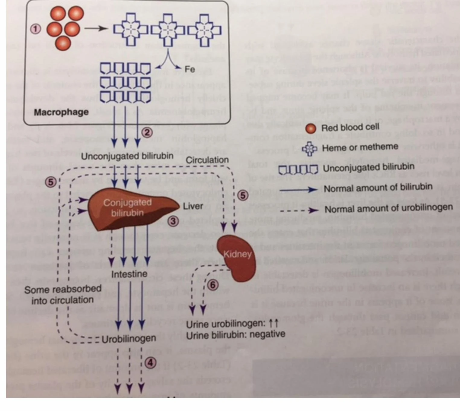
Inside the macrophage, the enzyme heme oxygenase breaks down the heme ring into biliverdin, which is then reduced to bilirubin. Bilirubin exists in two forms:
Unconjugated (Indirect) Bilirubin: Hydrophobic and must bind to albumin for transport to the liver.
Conjugated (Direct) Bilirubin: Created in the liver by adding two glucuronic acid molecules, making it water-soluble.
This diagram illustrates the metabolic pathway of bilirubin, starting from the breakdown of red blood cells in macrophages, its conjugation in the liver, and its subsequent excretion or reabsorption. It also shows the levels of urobilinogen and bilirubin in urine.
2. Intravascular Hemolysis (Fragmentation)
The remaining 10% of destruction occurs within the blood vessels due to mechanical trauma (e.g., valve narrowing) or enzyme defects. This results in the release of free hemoglobin directly into the plasma.
Characteristic findings of intravascular hemolysis include:
Hemoglobinemia: Free hemoglobin in the plasma.
Hemoglobinuria: Hemoglobin excreted in the urine.
Schistocytes: Fragmented red blood cells visible on a blood smear.
Exam Tip: Haptoglobin Levels
A significantly decreased haptoglobin level is one of the most reliable indicators of intravascular hemolysis, as haptoglobin is consumed while binding to free hemoglobin.
Iron Salvage and Oxidation Prevention
To prevent oxidative damage from free hemoglobin and to recycle precious iron, the body utilizes a three-step salvage system:
Haptoglobin: Binds to free hemoglobin dimers in the plasma, forming a complex that is transported to macrophages for recycling.
Hemopexin: If hemoglobin is not bound by haptoglobin, it may oxidize into methemoglobin. Hemopexin then binds to the released metheme to salvage the iron.
Methemalbumin: If the hemopexin system becomes saturated, metheme binds to albumin. Albumin acts as a temporary carrier until hemopexin becomes available to transfer the metheme to the liver.
Iron Recycling
The body is highly efficient at recycling iron. While we ingest iron through meat, eggs, and leafy greens, most of the iron used for new hemoglobin comes from the recycling of senescent RBCs. Iron is stored in the liver and bone marrow as ferritin (readily available) or hemosiderin (less readily available).
Summary of Laboratory Findings
When hemolysis occurs, the bone marrow attempts to compensate by increasing production, leading to reticulocytosis (increased reticulocyte count).
Finding
Extravascular (Macrophage)
Intravascular (Fragmentation)
Haptoglobin
Normal or slightly ↑
↓↓ (Decreased)
Bilirubin
↑ (Unconjugated/Direct)
↑ (Unconjugated)
Hemoglobin
Normal in plasma
↑ in plasma/urine
RBC Morphology
Spherocytes often present
Schistocytes often present
LDH
↑
↑
Keep reading for free.
Create an account to finish this reading and make it part of your study routine.