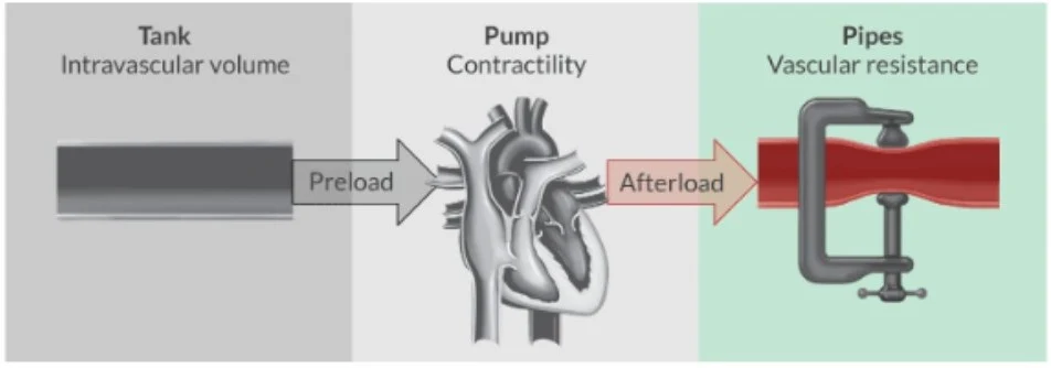
Hemodynamics is the study of how blood moves through the cardiovascular system, governed by the fundamental principles of fluid dynamics. To master this topic, it is helpful to view the cardiovascular system through the "Tank, Pump, and Pipes" model: the heart acts as the pump, the blood vessels act as the pipes, and the blood volume acts as the tank. Understanding the interrelationship between these variables is essential for managing patient stability and oxygen delivery.
The Physics of Flow: Ohm’s and Poiseuille’s Laws
The foundation of hemodynamic understanding is Ohm’s Law, which relates flow to the pressure gradient and resistance. In a clinical setting, we express this relationship to calculate Mean Arterial Pressure (MAP):
MAP=80(CO×SVR)+CVP
In this equation, Cardiac Output (CO) is the flow, Systemic Vascular Resistance (SVR) is the resistance, and the difference between MAP and Central Venous Pressure (CVP) represents the pressure gradient.
While Ohm's Law provides the macro-view, Poiseuille’s Law refines our understanding by incorporating the physical properties of the vessels themselves:
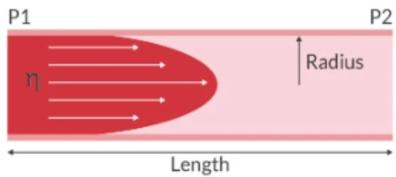
Q=8ηLπR4ΔP
Where:
Q is blood flow
R is vessel radius
ΔP is the pressure gradient
η is blood viscosity
L is vessel length
Because the radius (R) is raised to the fourth power, even minute changes in vessel diameter—particularly in the arterioles—have a profound impact on blood flow.
Diagram illustrating fluid flow in a tube, showing velocity profile and indicating length and radius.
Blood Flow Types and Viscosity
Blood flow can be categorized as laminar (smooth, streamlined flow), turbulent (irregular, chaotic flow), or transitional. Additionally, viscosity (η) plays a critical role in resistance. Viscosity is primarily influenced by hematocrit and temperature; for instance, higher temperatures or lower hematocrit levels decrease viscosity, thereby improving flow.
The Pump: Cardiac Output and Contractility
Cardiac Output (CO) is the volume of blood the heart pumps per minute. It is the product of Heart Rate (HR) and Stroke Volume (SV). The efficiency of this "pump" is determined by three primary factors: preload, contractility, and afterload.
Contractility (Inotropy)
Contractility, or inotropy, refers to the innate ability of myocardial sarcomeres to perform work. Unlike preload or afterload, contractility is an intrinsic property of the muscle that is independent of the volume of blood in the chamber. This process is heavily dependent on the availability of calcium.
Excitation-Contraction Coupling
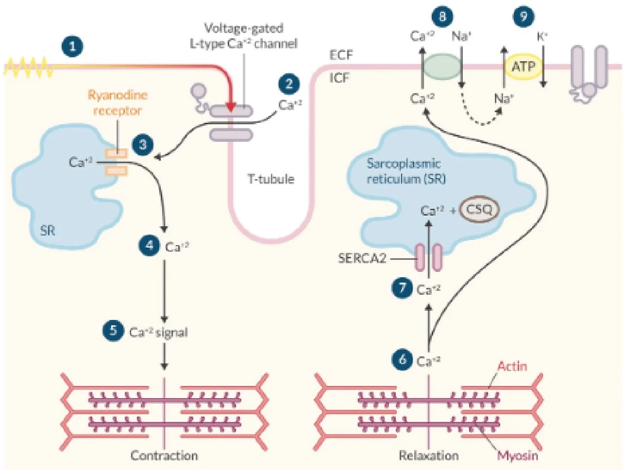
The mechanism by which an electrical signal results in mechanical contraction is known as excitation-contraction coupling. When an action potential depolarizes the cell, it opens L-type calcium channels, allowing calcium to enter the myocyte. This trigger leads to calcium-induced calcium-release via the ryanodine receptor from the sarcoplasmic reticulum. The released calcium binds to troponin C, facilitating the formation of cross-bridges between actin and myosin filaments within the sarcomere.
Diagram illustrating the process of calcium handling in muscle cells, leading to contraction and relaxation. It shows the role of the sarcoplasmic reticulum, T-tubule, and various ion channels and transporters in regulating intracellular calcium levels.
Relaxation occurs when calcium is sequestered back into the sarcoplasmic reticulum by the SERCA2 pump or removed from the cell via the sodium/calcium exchanger (NCX).
The Tank: Preload and Ventricular Function
Preload represents the "tank" level—specifically, the ventricular wall tension at the end of diastole. It is influenced by total blood volume, venous tone, and the atrial kick (the final contribution of atrial contraction to ventricular filling).
The Frank-Starling Mechanism
The relationship between preload and stroke volume is described by the Frank-Starling mechanism. This principle states that as preload increases, the myocardial fibers are stretched, leading to an increase in stroke volume up to an optimal stretch point. However, if the sarcomeres are overstretched beyond this point, the force of contraction decreases, and output falls.
Diagram illustrating the effect of stretching on muscle contraction, showing three states: under-stretched, optimally stretched, and over-stretched, with explanations for each.
Exam Tip: Remember that ventricular compliance (the ease with which the ventricle expands) is a key determinant of preload. A "stiff" ventricle has low compliance, which can impair filling and reduce preload.
The Pipes: Afterload and Systemic Vascular Resistance
Afterload is the resistance the heart must pump against to eject blood into the systemic circulation. This is primarily determined by Systemic Vascular Resistance (SVR).
Diagram illustrating the relationship between intravascular volume (tank), pump contractility (heart), and vascular resistance (pipes) in the circulatory system, with preload and afterload as key factors.
Changes in afterload have significant implications for myocardial oxygen demand. For example, hypertension increases afterload, forcing the myocardium to generate higher pressures to open the aortic valve, which in turn increases oxygen consumption.
Summary of Hemodynamic Interrelationships
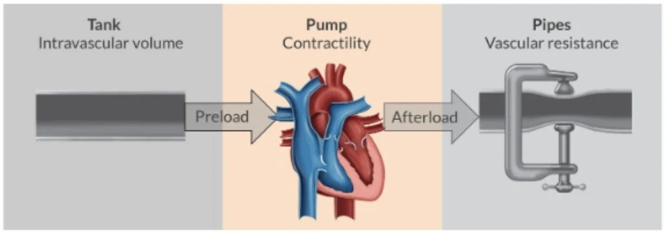
To visualize how these variables interact, we can use the following model:
Diagram illustrating the relationship between intravascular volume (preload), pump contractility (heart), and vascular resistance (afterload) in the circulatory system.
In summary, maintaining adequate oxygen delivery (DO2) requires a balance between the "pump" (Cardiac Output), the "tank" (Oxygen content in the blood), and the "pipes" (the ability of the vessels to maintain pressure and perfusion).
Keep reading for free.
Create an account to finish this reading and make it part of your study routine.