Anemia: Classification and Diagnosis
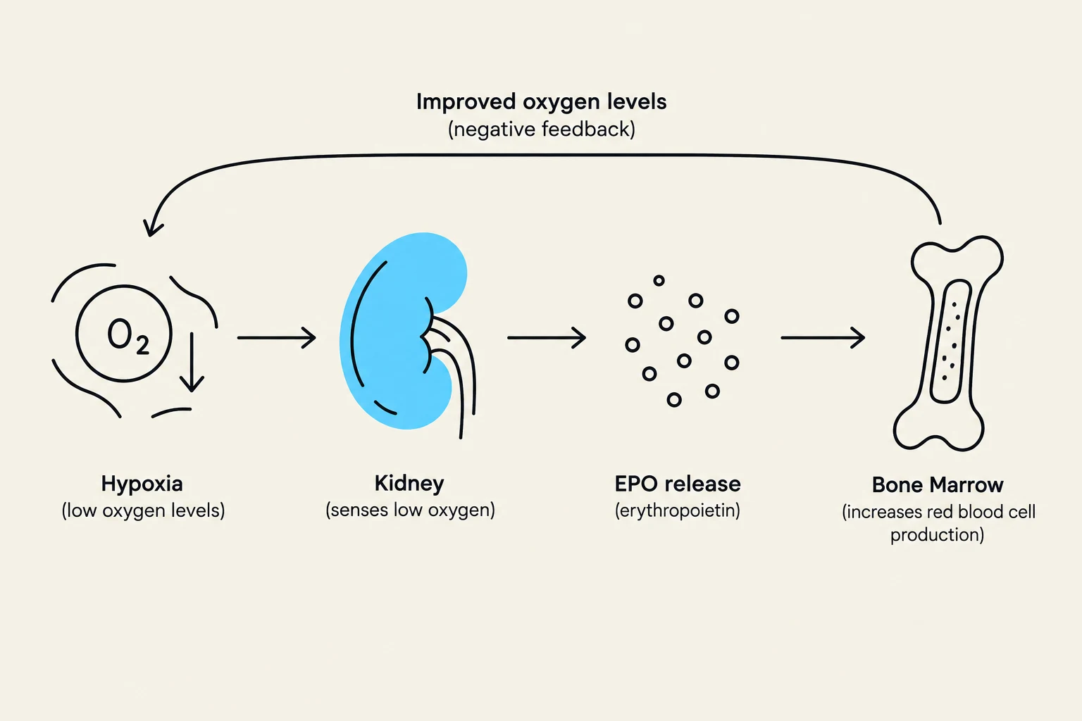
Anemia is defined by a decrease in hemoglobin (Hb) or RBC mass, leading to reduced oxygen-carrying capacity. To diagnose the underlying cause, clinicians rely on the Mean Corpuscular Volume (MCV) to classify the anemia:
1. Microcytic Anemia (Low MCV)
Typically caused by Iron Deficiency Anemia (IDA) or thalassemia.
- IDA Profile: Characterized by ↓ ferritin, ↓ serum iron, and ↑ Total Iron Binding Capacity (TIBC).
- Anemia of Chronic Inflammation: Unlike IDA, this presents with ↓ iron but normal or ↑ ferritin because iron is sequestered in storage rather than being depleted.
2. Macrocytic Anemia (High MCV)
Caused by impaired DNA synthesis, most commonly due to Vitamin B12 (cobalamin) or folate deficiency.
- Critical Distinction: B12 deficiency presents with neurologic symptoms (e.g., paresthesia, confusion), whereas folate deficiency does not.
- Warning: Treating a B12-deficient patient with only folate can correct the cell size (masking the anemia) while allowing irreversible neurological damage to progress.
3. Normocytic Anemia (Normal MCV)
To differentiate causes of normocytic anemia, clinicians use the reticulocyte count (immature RBCs):
- High Reticulocyte Count: Suggests the marrow is responding to blood loss or hemolysis.
- Low Reticulocyte Count: Suggests a production failure, such as chronic kidney disease (CKD) or bone marrow issues.
Exam Tip: If a patient has macrocytic anemia, always check both B12 and folate levels. Replacing folate alone can "mask" a B12 deficiency by fixing the RBC size while the neurological damage continues.
Hemostasis: The Clotting Process
Hemostasis is the arrest of bleeding and occurs in two distinct, overlapping stages:
Primary Hemostasis (Platelet Plug)
When a vessel is injured, the subendothelium (collagen) is exposed. Von Willebrand factor (vWF) acts as "glue," allowing platelets to adhere to the site. Platelets then undergo aggregation via fibrinogen and the GP IIb/IIIa receptor to form a temporary platelet plug. Deficiencies in this stage (e.g., vWD or low platelets) typically cause superficial bleeding, such as petechiae or mucosal bleeding.
Secondary Hemostasis (Coagulation Cascade)
This stage involves a cascade of clotting factors that stabilize the platelet plug into a hard clot:
- Initiation/Propagation: Tissue Factor (TF) and Factor VIIa activate the cascade, eventually leading to the activation of Factor X.
- Thrombin Generation: Factor Xa (along with Va, Ca2+, and phospholipids) forms the prothrombinase complex, which converts Prothrombin (II) into Thrombin (IIa).
- Fibrin Formation: Thrombin converts Fibrinogen into Fibrin.
- Stabilization: Factor XIII cross-links the fibrin monomers to create a stable, insoluble mesh.
Deficiencies in these factors (e.g., Hemophilia) result in deep bleeding, such as hemarthrosis (bleeding into joints).
Fibrinolysis and Monitoring
To prevent excessive clotting, the body uses fibrinolysis. The enzyme tPA converts plasminogen into plasmin, which breaks down fibrin clots. The breakdown products of this process are measured as the D-dimer test, which is useful in diagnosing conditions like DVT or PE.
Clinical monitoring of the cascade is done via:
- PT/INR: Monitors the extrinsic pathway (e.g., for Warfarin).
- aPTT: Monitors the intrinsic pathway (e.g., for Heparin).
Thrombosis and Pharmacology
Thrombosis risk is summarized by Virchow’s Triad: stasis (immobility), endothelial injury (trauma/inflammation), and hypercoagulability (cancer/genetic states).
Antiplatelets (Arterial Focus)
Arterial thrombosis is primarily platelet-driven. Drugs used here include:
- Aspirin (ASA): An irreversible COX inhibitor that reduces Thromboxane A2.
- Clopidogrel: A P2Y12 ADP receptor antagonist used for coronary stents.
Anticoagulants (Venous Focus)
Venous thrombosis is primarily fibrin-driven.
- Unfractionated Heparin (UFH): Potentiates antithrombin to inhibit both Xa and IIa. It has a rapid onset and is used in urgent situations. It is monitored via aPTT and can be reversed with Protamine.
- Calculation Note: 1 mg of protamine reverses 100 units of UFH.
- Low Molecular Weight Heparin (LMWH): (e.g., Enoxaparin) Primarily inhibits Factor Xa. It has a longer half-life and more predictable dosing than UFH.
- Warfarin: A Vitamin K antagonist that inhibits factors II, VII, IX, and X. It has a slow onset and requires PT/INR monitoring. It is reversed by Vitamin K.
- Direct Oral Anticoagulants (DOACs):
- Direct Thrombin Inhibitors: Dabigatran inhibits Factor IIa directly. Reversed by Idarucizumab.
- Direct Factor Xa Inhibitors: Rivaroxaban and Apixaban directly inhibit Factor Xa without needing antithrombin. Reversed by Andexxa.
Disseminated Intravascular Coagulation (DIC)
DIC is a life-threatening paradox where widespread activation of coagulation leads to the consumption of all platelets and clotting factors. This results in a state where the patient simultaneously experiences thrombosis (microthrombi causing organ ischemia) and bleeding (due to lack of remaining factors).
Summary Table for Quick Review
| Drug | Target | Monitoring | Antidote |
|---|
| UFH | Antithrombin → Xa + IIa | aPTT | Protamine |
| Warfarin | ↓ Vit K → II, VII, IX, X | PT/INR | Vitamin K |
| Dabigatran | Direct IIa | None | Idarucizumab |
| Apixaban | Direct Xa | None | Andexxa |
| Aspirin | COX → ↓ TXA2 | None | N/A |