Hematology: Anemia, Leukemia, and Blood Cell Function
An overview of hematology, covering the study of blood, its components, and disorders such as anemia and leukemia. Explains red blood cell function, anemia etiologies, sickle cell anemia, and diagnostic tests like hematocrit and white blood cell counts.
Hematology is the branch of medicine dedicated to the study of blood, its components, and the various physiological functions they perform within the body. To understand hematological disorders, one must first understand the primary constituents of blood: Red Blood Cells (RBCs), which transport oxygen; White Blood Cells (WBCs), which serve as the body's defense mechanism; and Thrombocytes (platelets), which are essential for clotting.
Understanding Anemia and Its Etiologies
It is a common misconception that anemia is a primary disease. In clinical practice, anemia is viewed as a condition resulting from various underlying etiologies rather than a standalone diagnosis. Essentially, anemia occurs when there is a deficiency in the body's ability to transport oxygen effectively, which can be categorized into three main pathophysiological causes:
Lack of Production: Often described as "manufacturing problems," this occurs when the body cannot produce an adequate number of RBCs.
Destruction of RBCs: This is known as hemolysis, where red blood cells are destroyed faster than they can be replaced.
Insufficient Quantity: A general state where there are simply not enough RBCs circulating in the bloodstream.
This diagram illustrates various types of anemias, including hypochromic anemia due to iron or vitamin deficiency, pernicious anemia caused by a lack of intrinsic factor, aplastic anemia from malfunctioning bone marrow, sickle cell anemia, and secondary anemia linked to bleeding or chronic diseases. It also depicts erythroblastosis fetalis, where antibodies destroy red blood cells.
Sickle Cell Anemia and Vasoocclusive Crisis
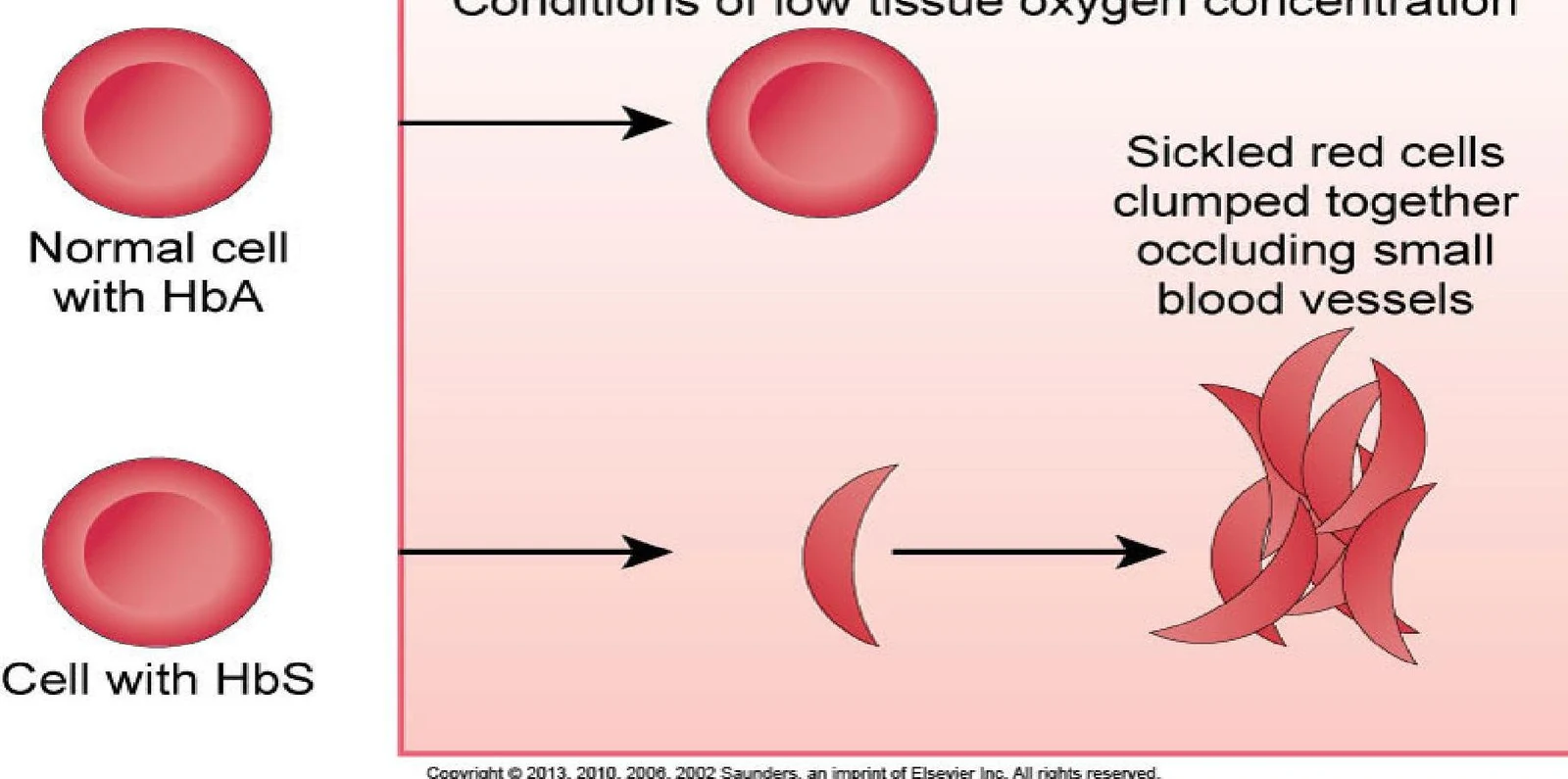
Sickle cell anemia serves as a primary exemplar of an inherited red blood cell disorder. In this condition, the red blood cells lose their flexible, biconcave shape and deform into a rigid "sickle" shape.
This illustration depicts the difference between normal red blood cells with HbA and sickle-shaped red blood cells with HbS, showing how the latter can clump together and obstruct blood vessels under low oxygen conditions.
This deformation leads to a life-threatening event known as a Vasoocclusive Crisis. During a crisis, these clumped, sickled cells obstruct blood vessels, leading to ischemia—a restriction in blood supply to tissues, causing a shortage of oxygen. The consequences of this obstruction can be devastating and include:
Organ Damage: Ischemia can lead to infarction (tissue death) in the kidneys, the brain (resulting in a stroke), or the spleen.
Hand/Foot Syndrome: Acute pain and swelling in the hands and feet.
Respiratory Distress: Obstruction can cause acute chest pain, fever, and hypoxia.
Clinical Management Tip: The "HOP" Approach
When managing a sickle cell crisis, focus on three pillars:
Hydration and Electrolytes
Oxygenation and Bed Rest
Pain Relief
Laboratory Monitoring and Diagnostics
To monitor hematological health, clinicians rely on specific laboratory tests to evaluate the concentration and type of blood cells present.
Hematocrit (Hct)
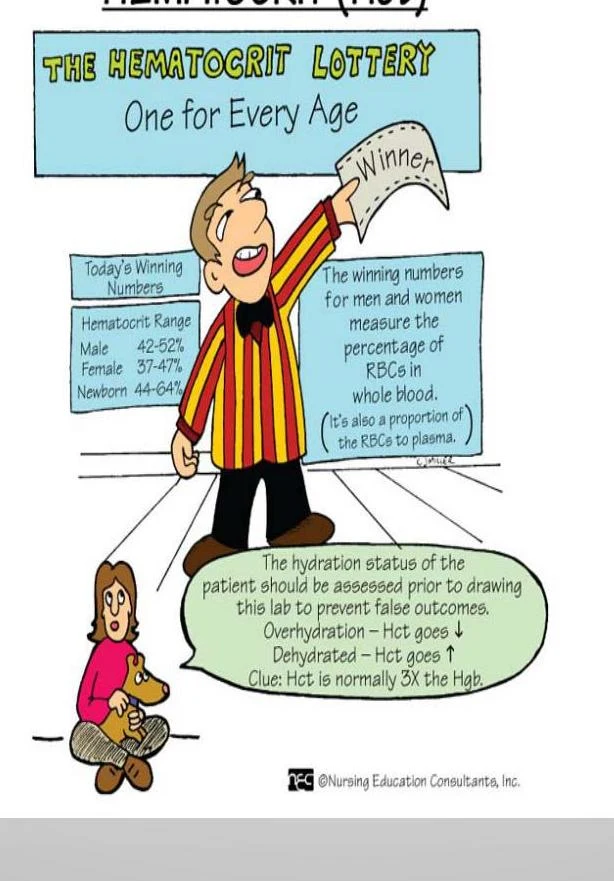
The Hematocrit measures the percentage of the total blood volume that is occupied by red blood cells.
This illustration explains hematocrit ranges for different age groups and genders, and how hydration status can affect hematocrit levels. It also defines hematocrit as the percentage of red blood cells in whole blood and its relation to plasma.
Critical Note: Always consider the patient's hydration status when interpreting Hct. If a patient is dehydrated, the Hct may appear falsely elevated because there is less plasma volume relative to the number of RBCs.
White Blood Cell (WBC) Count
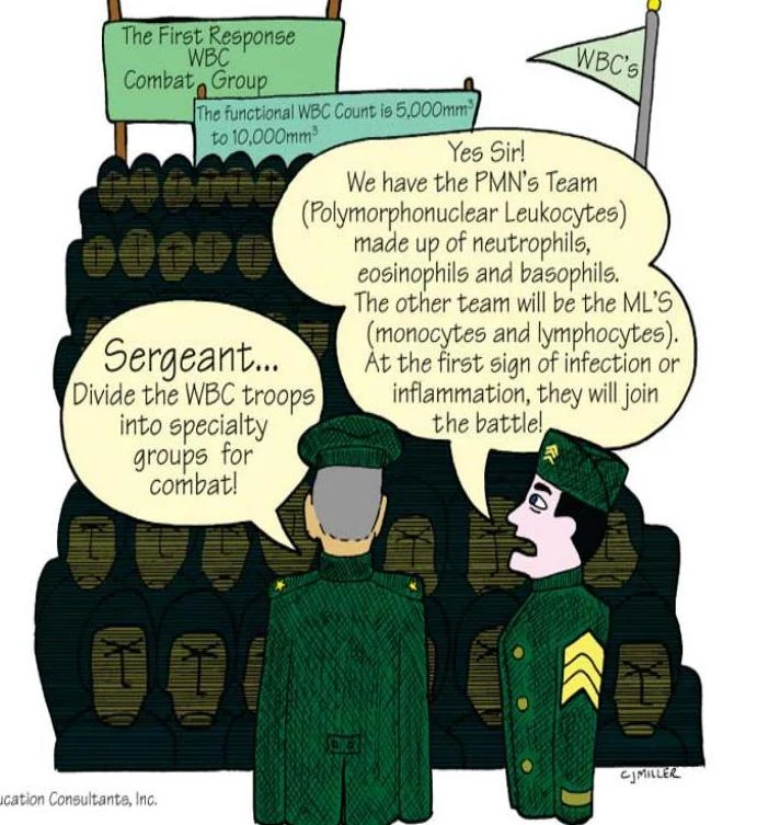
The WBC count evaluates the body's immune response. Leukocytes are generally categorized into two functional "teams":
Polymorphonuclear Leukocytes (PMNs): This group includes neutrophils, eosinophils, and basophils.
Monocytes and Lymphocytes (MCS): These cells are primary responders to signs of infection or inflammation.
This illustration depicts white blood cells (WBCs) as a combat group, with different types of WBCs like neutrophils, eosinophils, basophils, monocytes, and lymphocytes being organized into specialty groups to fight infection or inflammation. The normal functional WBC count is stated as 5,000 to 10,000 cells per cubic millimeter.
Leukemia: Uncontrolled Cellular Growth
While anemia involves issues with red blood cells, Leukemia is a malignancy characterized by the uncontrolled production of immature white blood cells within the bone marrow.
Because these cells are immature, they do not function correctly to fight infection, and their rapid production crowds out healthy blood cells in the bone marrow. Although the exact cause of leukemia remains unknown, several factors are believed to contribute to its development:
Genetic Factors: Inherited predispositions.
Environmental Factors: Exposure to ionizing radiation, certain chemicals, and drugs.
Immunologic Factors: Underlying issues with the immune system.
Clinical Correlation: In patients with leukemia, clinicians must closely monitor for signs of thrombocytopenia (low platelets) and neutropenia (low neutrophils), as these significantly increase the risk of bleeding and infection, respectively.
Keep reading for free.
Create an account to finish this reading and make it part of your study routine.