


To understand how the heart functions as a pump, one must first understand its physical architecture. The heart is not merely a single muscle; it is a complex, multi-layered organ encased in a protective sac, designed to facilitate the continuous flow of blood through the pulmonary and systemic circuits. Understanding the layers of the heart and the protective pericardium is fundamental to recognizing how various diseases—such as inflammation or infection—impact cardiac output and patient stability.
The Pericardial Sac
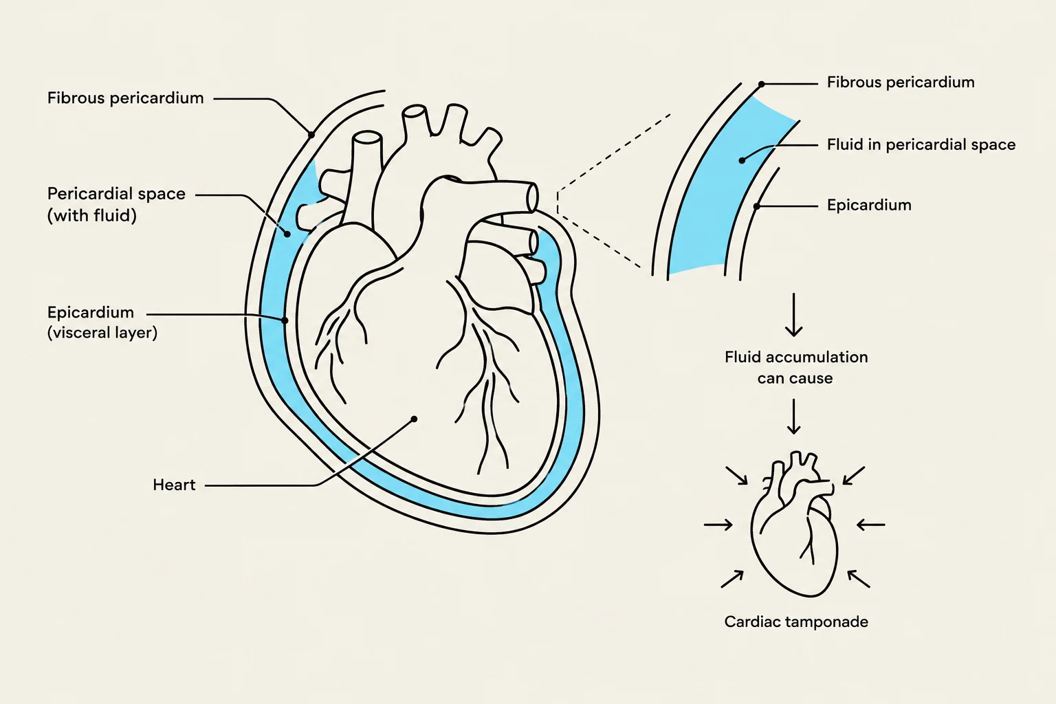
The heart is enclosed within a protective structure known as the pericardial sac. This sac serves to anchor the heart within the chest and provides a layer of protection against friction and infection.
When the surfaces within this sac become inflamed, a condition known as pericarditis can occur. Pericarditis is characterized by inflammation of the pericardium, which can lead to reduced ventricular filling, subsequently decreasing cardiac output and blood pressure.
Clinical Note: Pericardial Friction Rub During a physical examination, a clinician may hear a "pericardial friction rub." This occurs when the inflamed pericardial surfaces rub together during the heartbeat.
If fluid accumulates excessively within this sac, it can lead to a life-threatening complication called cardiac tamponade, which requires immediate medical intervention, such as a pericardiocentesis (the removal of fluid from the area).
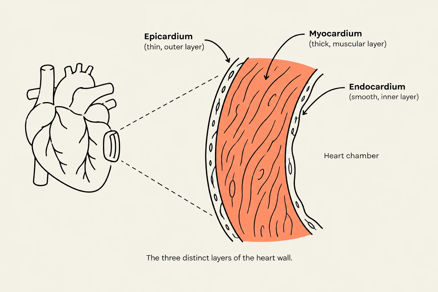
The Three Cardiac Layers
Keep reading for free.
Create an account to finish this reading and make it part of your study routine.
Continue reading freeAlready have an account? Log in
Updated