This comprehensive guide covers the anatomy, mechanics, and developmental considerations of the respiratory system, along with methods for physical assessment and interpretation of findings.
The respiratory system is vital for life, serving not only to supply oxygen for energy production but also to remove carbon dioxide and maintain the body's acid-base balance. A thorough health assessment requires a deep understanding of the anatomical landmarks, the mechanics of breathing, and how these functions shift as a person moves through different stages of life. By mastering these concepts, clinicians can effectively differentiate between normal physiological findings and signs of underlying pathology.
Anatomy and Thoracic Landmarks
To perform an accurate physical exam, a clinician must be able to map the internal structures of the lungs onto the external surface of the chest. The thorax contains the mediastinum, which houses the heart, trachea, and esophagus, and the pleural cavities, which hold the lungs. The lungs themselves are protected by two layers of membranes: the visceral pleura, which lines the lungs, and the parietal pleura, which lines the chest wall and diaphragm.
Anterior and Posterior Landmarks
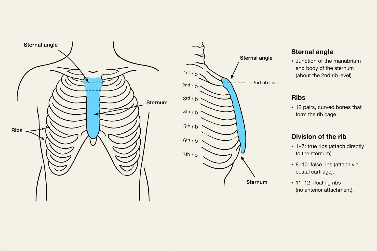
Assessment begins with identifying key bony landmarks. On the anterior chest, the suprasternal notch is the U-shaped hollow above the sternum. A critical landmark is the sternal angle (Angle of Louis), where the manubrium meets the body of the sternum. This angle marks the level of the tracheal bifurcation (T4 level) and serves as the starting point for counting the second rib.
On the posterior chest, the vertebra prominens (C7) is the most prominent spinous process when the head is flexed. While spinous processes align with their corresponding ribs down to T4, below that level, each process overlies the rib below.
Reference Lines and Lung Lobes
Clinicians use vertical reference lines to locate findings precisely:
Midsternal/Midspinal: Along the center of the chest or spine.
Midclavicular: Running vertically through the midpoint of the clavicles.
Axillary lines: Located on the lateral aspect of the thorax (anterior, mid, and posterior).
The lungs are not symmetrical. The right lung has three lobes (RUL, RML, RLL) divided by the oblique and horizontal fissures. The left lung has only two lobes (LUL, LLL) due to the space occupied by the heart. Because the lobes sit in diagonal, sloping segments rather than horizontal layers, the anterior chest primarily allows access to the upper and middle lobes, while the posterior chest provides access to almost all of the lower lobes.
An illustration showing the sternal angle and the division of the ribs into true, false, and floating ribs.
Respiratory Mechanics and Control
Respiration is a complex process regulated by the brainstem, specifically the pons and medulla. The primary driver for breathing is humoral regulation, where an increase in carbon dioxide levels in the blood (hypercapnia) acts as the main stimulus to breathe.
The Mechanics of Breathing
Breathing is divided into two distinct phases:
Inspiration: An active process. The diaphragm contracts and descends, increasing the vertical diameter of the chest, while the intercostal muscles lift the ribs to increase the anteroposterior (AP) diameter.
Expiration: A mostly passive process. As the diaphragm relaxes and domes upward, the natural elastic recoil of the lung tissue pushes air out.
In cases of respiratory distress, the body may recruit accessory muscles (such as the sternocleidomastoid, scalene, or trapezius) to assist with inspiration, or abdominal muscles to assist with forced expiration.
Developmental Considerations Across the Lifespan
Respiratory function is not static; it changes significantly from birth through old age.
Infants and Children
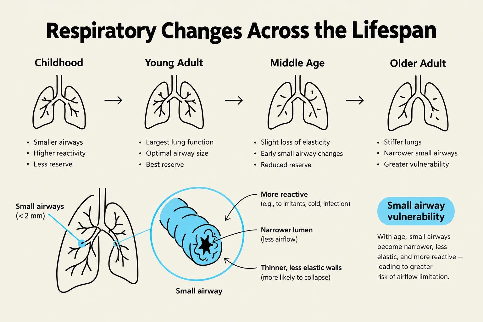
The respiratory system is unique because it is the only system that does not function until birth. Children have smaller, immature airways and significantly fewer alveoli (approximately 70 million at birth compared to 300 million in an adult). This makes them highly vulnerable to smoke exposure and lower respiratory infections.
Pregnancy
During pregnancy, the enlarging uterus elevates the diaphragm by approximately 4 cm. This causes the transverse diameter of the chest to increase and the costal angle to widen. While tidal volume increases by about 40%, many pregnant people experience physiologic dyspnea (shortness of breath) due to these mechanical shifts, even without underlying lung disease.
Older Adults
Aging leads to a more rigid thoracic structure. Key changes include:
Calcification of costal cartilages: This reduces the mobility of the thorax.
Decreased elastic recoil: The lungs become less distensible.
Loss of alveolar septa: This reduces the surface area available for gas exchange.
These changes result in a decreased vital capacity and an increased residual volume. Consequently, older adults are at a higher risk for atelectasis (collapsed lung tissue) and infections because they may have a weaker cough and fewer airway reflexes.
This diagram illustrates the changes in respiratory function and airway characteristics across different life stages, from childhood to older adulthood, highlighting the increasing vulnerability of small airways with age.
Clinical Application: The Physical Exam
A complete respiratory assessment follows a systematic sequence: Inspection, Palpation, Percussion, and Auscultation.
Exam Tip: When auscultating, always compare side-to-side at each site. A normal breath sound heard in the wrong location—such as harsh bronchial sounds heard over the peripheral lung fields—is a major indicator of consolidation or pneumonia.
Interpreting Breath Sounds
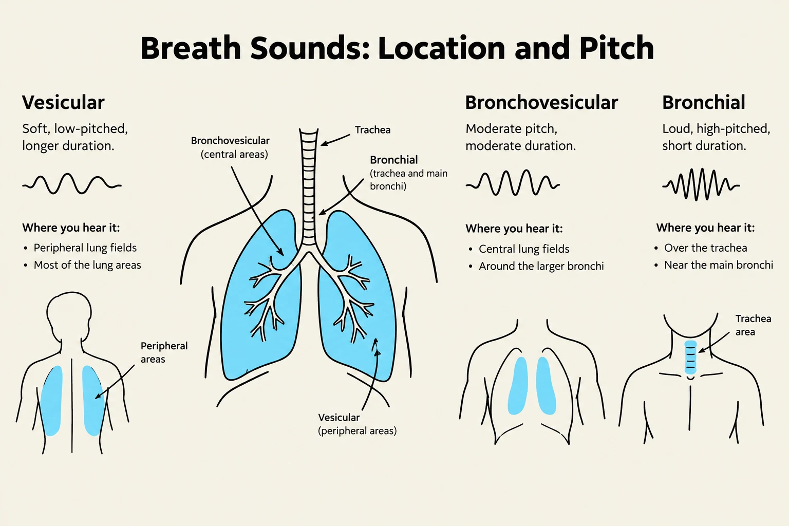
Normal Sounds: Include vesicular (soft, rustling sounds over peripheral lung fields), bronchovesicular (mixed pitch over major bronchi), and bronchial (loud, high-pitched sounds over the trachea).
Wheezes: Musical, squeaking sounds caused by narrowed airways (asthma, emphysema).
Stridor: A high-pitched, crowing sound indicating upper airway obstruction; this is a medical emergency.
Worked Example: Assessing a Patient with Chronic Symptoms
Consider a patient presenting with a chronic morning cough and a "barrel chest" (where the AP diameter equals the transverse diameter).
If the clinician notes hyperresonance on percussion and decreased tactile fremitus, these findings, combined with a history of heavy smoking, strongly suggest emphysema. In emphysema, air is trapped in the lungs, leading to the characteristic barrel chest and hyperresonant percussion notes.
This illustration details the location and pitch of different breath sounds: vesicular, bronchovesicular, and bronchial. It shows where these sounds are typically heard in the respiratory system.
Keep reading for free.
Create an account to finish this reading and make it part of your study routine.