An educational guidebook describing immunohistochemistry as a precise immunoassay used to determine the expression of biomarkers in tissue for cancer diagnosis and classification. It covers pre-analytical factors, antigen retrieval, antibody selection, detection systems, visualization, and post-analytical interpretatio
Immunohistochemistry (IHC) is a sophisticated immunoassay used to achieve the microscopic localization of specific antigens within tissue sections. By utilizing antibodies labeled with either pigmented material or fluorescent dyes, pathologists can identify the presence, location, and abundance of specific biomarkers. This technique is a cornerstone of modern pathology, serving as a vital tool for the differential diagnosis, classification, and prognostic assessment of various cancers and infectious diseases.
While IHC shares fundamental principles with the Enzyme-Linked Immunosorbent Assay (ELISA), it is uniquely applied to the complex, three-dimensional architecture of tissue sections. To ensure clinical reliability, the field is moving away from treating IHC as a simple "special stain" and toward a rigorous, quantifiable immunoassay framework.
The "Total Test" Concept and Standardization
Historically, IHC has been plagued by poor reproducibility, often due to inconsistencies in how tissues were handled or variations in antibody quality. To combat this, modern laboratories adopt the "Total Test" approach. This concept posits that standardization must be enforced across every single phase of the process—from the moment a biopsy is taken to the final digital image analysis.
Standardization is not merely a laboratory concern; it begins in the surgery room. If a tissue sample is not fixed correctly at the source, no amount of advanced laboratory technology can recover the lost antigen information.
Exam Tip: When discussing IHC reliability, always emphasize that the "Total Test" includes pre-analytical, analytical, and post-analytical phases. A failure in any one phase compromises the entire result.
Phase I: Pre-analytical Factors
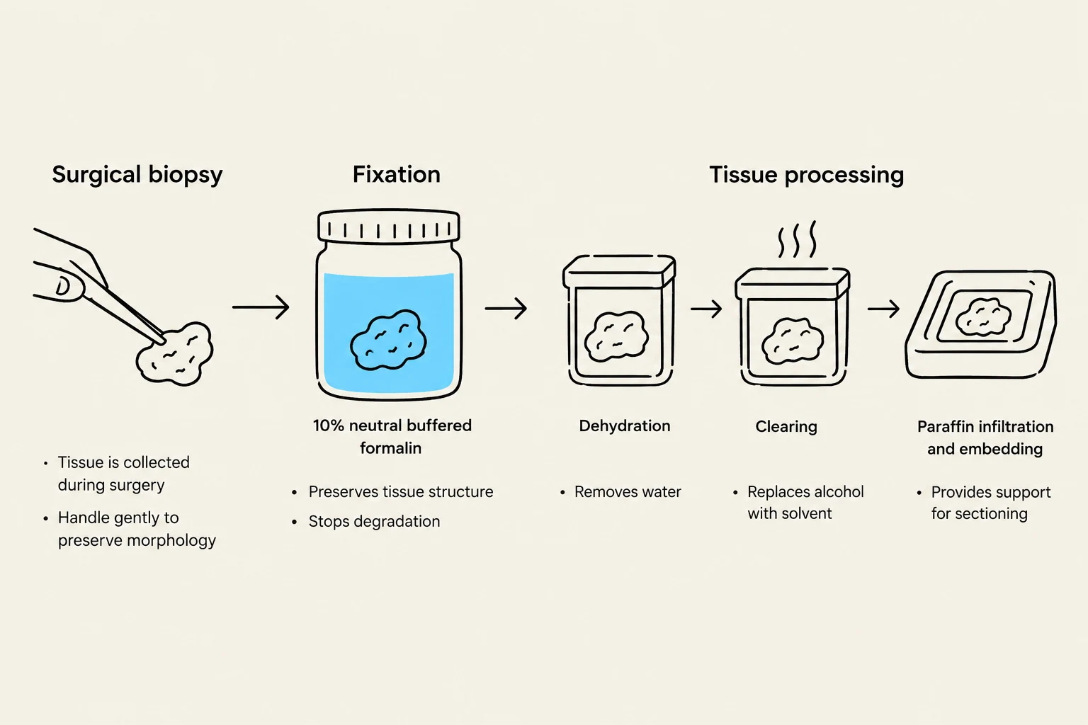
The pre-analytical phase is arguably the most critical for preserving antigen integrity. Most IHC is performed on Formalin-Fixed, Paraffin-Embedded (FFPE) tissue. This process involves several delicate steps:
Biopsy and Accessioning: The initial collection and identification of the specimen.
Grossing: The macroscopic examination and slicing of the tissue.
Fixation: The use of chemical fixatives (typically formalin) to preserve tissue structure.
Processing and Embedding: Dehydrating the tissue and infiltrating it with paraffin wax to create a stable block.
Sectioning: Cutting the block into extremely thin slices, typically ∼4\mum thick.
Variables such as the delay between surgery and fixation (ischemic time), the type of fixative used, and the dehydration protocols can significantly impact the availability of epitopes.
This diagram illustrates the steps involved in processing tissue samples, from surgical biopsy to paraffin infiltration and embedding, for histological analysis.
Phase II: The Analytical Process
The analytical phase is where the actual "staining" occurs. This involves a series of highly optimized chemical reactions designed to unmask antigens and visualize them.
Antigen Retrieval
During fixation, proteins often form cross-links that "mask" the epitopes (the specific parts of the antigen the antibody recognizes). To reveal these epitopes, laboratories use antigen retrieval techniques:
Heat-Induced Epitope Retrieval (HIER): Using heat (via pressure cookers, autoclaves, or microwave ovens) in a specific buffer solution.
Proteolytic (Enzymatic) Retrieval (PIER): Using enzymes like pepsin to digest the protein matrix and expose the antigens.
Antibody Selection and Detection Systems
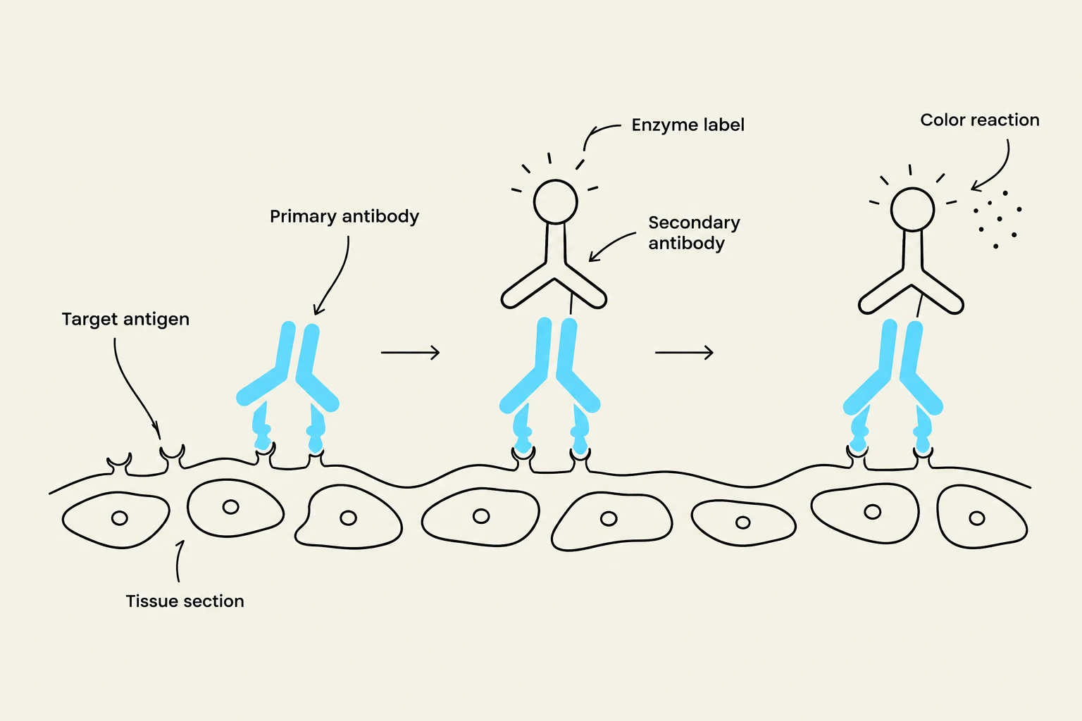
The specificity of the test relies on the primary antibody, which is selected to bind specifically to the target antigen. These antibodies can be monoclonal (highly specific, derived from a single cell line) or polyclonal (a mixture of antibodies that recognize different parts of the same antigen).
Once the primary antibody is bound, a secondary antibody is applied. Modern detection systems often use polymer-based detection, which links multiple enzyme molecules to the secondary antibody to significantly increase sensitivity and reduce background noise compared to older avidin-biotin methods.
Visualization
To see the results under a microscope, a chromogen (such as DAB, which produces a brown precipitate) is used to create a visible color change at the site of the antigen-antibody complex. Finally, a counterstain (like hematoxylin) is applied to reveal the overall tissue architecture, allowing the pathologist to see where the specific staining is occurring relative to the cell nuclei and cytoplasm.
Diagram illustrating the process of antibody-based detection of a target antigen in a tissue section, involving a primary antibody, a secondary antibody with an enzyme label, and a resulting color reaction.
Phase III: Post-analytical Interpretation
The final phase involves the pathologist interpreting the stained slides using bright-field microscopy. For many biomarkers, the result is not a simple "yes/no" but a semi-quantitative score.
For example, in companion diagnostics—where the IHC result determines if a patient is eligible for a specific targeted therapy—markers like HER2, ER (Estrogen Receptor), or PR (Progesterone Receptor) are scored on a scale (e.g., 0 to $3+$).
0 or 1+: Generally considered negative.
2+: Equivocal (may require further testing like FISH).
3+: Positive (indicating high expression of the biomarker).
Quality Assurance and the Future of IHC
To maintain high standards, laboratories participate in External Quality Assurance (EQA) schemes, such as NordiQC or UK NEQAS. These programs provide standardized materials to laboratories to monitor staining quality and ensure results are consistent across different institutions.
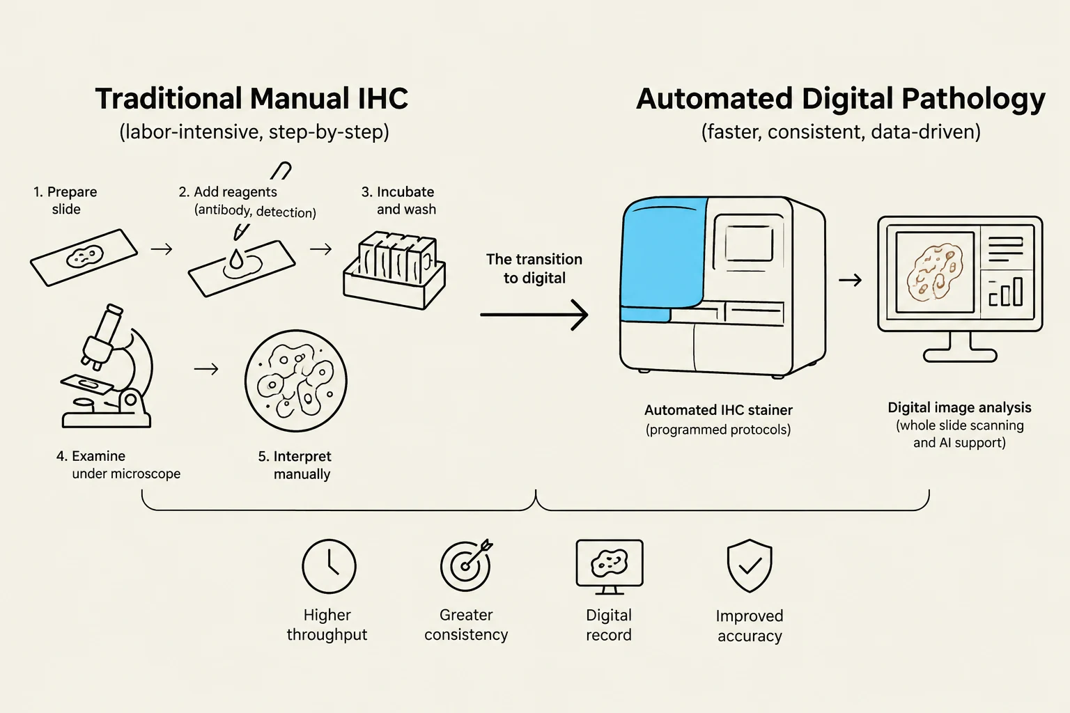
The field is also rapidly evolving through automation and digital pathology. Automated stainers increase reproducibility by removing human error from the timing and reagent application steps, while digital imaging and algorithms allow for more objective, quantifiable image analysis.
This diagram illustrates the process of immunohistochemistry (IHC), comparing traditional manual methods with automated digital pathology, highlighting the steps involved and the benefits of automation such as higher throughput, greater consistency, digital records, and improved accuracy.