


Understanding the human lower limb requires an integrated view of how bones, muscles, and connective tissues work in concert to facilitate movement, stability, and weight-bearing. This study article examines the organizational principles of limb compartments, the specific muscle groups of the thigh and gluteal region, and the critical skeletal structures of the pelvis, hip, and knee.
Organizational Principles: Compartments and Fascia
The muscles of the limbs are not merely scattered throughout the flesh; they are organized into functional groups known as limb compartments. These compartments are defined by deep fascia, a thick and inelastic layer of dense connective tissue.
To create these distinct spaces, the deep fascia dives between muscle groups to form intermuscular septa. These septa anchor directly into the periosteum (the outer layer) of the bones, effectively partitioning the limb into sections. This organization is highly efficient: muscles within a single compartment typically share the same primary actions, the same blood supply, and the same innervation via shared neurovascular bundles.
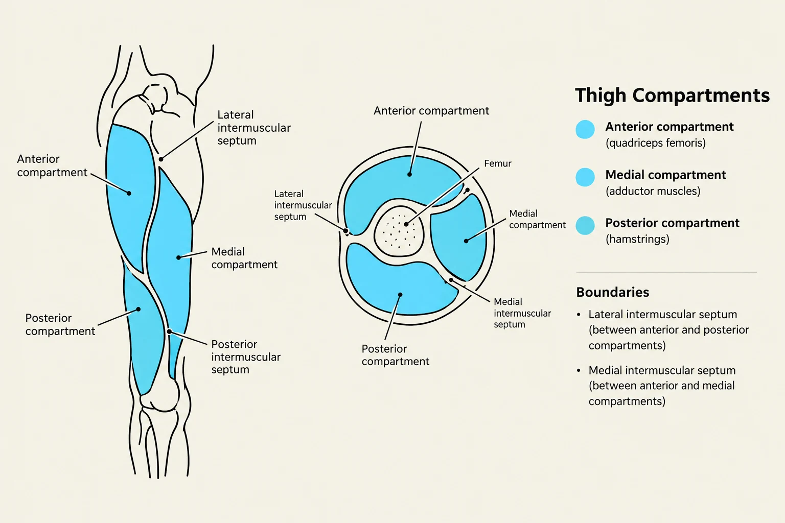
Exam Tip: If a question asks about the functional relationship between muscles in a compartment, remember that they are grouped by shared innervation and action due to this compartmentalization.
Muscular Anatomy of the Thigh
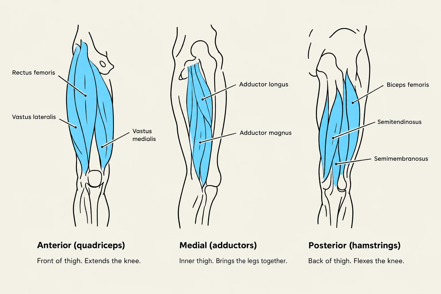
The thigh is divided into three primary compartments, each serving a distinct mechanical purpose in locomotion.
The Anterior Compartment
The primary occupants of the anterior compartment are the quadriceps femoris. This group consists of four muscles: the rectus femoris, vastus lateralis, vastus intermedius, and vastus medialis. Collectively, these muscles are responsible for knee extension, which is essential for activities like walking, kicking, or standing up from a seated position.
Keep reading for free.
Create an account to finish this reading and make it part of your study routine.
Continue reading freeAlready have an account? Log in
Updated