An overview of bone classification by shape, gross anatomy including compact and spongy bone, bone membranes, marrow types, and microscopic anatomy with bone cell types.
Bone is a specialized, vascularized connective tissue characterized by a mineralized matrix. Far from being static structures, bones serve critical roles in the body: they provide structural support for soft organs, protect vital structures like the brain and spinal cord, act as levers for movement, and serve as reservoirs for minerals like calcium and phosphorus. Additionally, bones are the site of hematopoiesis, the production of blood cells.
To understand how bones function and heal, one must distinguish between their various shapes and their internal histological organization.
Classification of Bones by Shape
Bones are categorized into five distinct groups based on their physical dimensions and complexity:
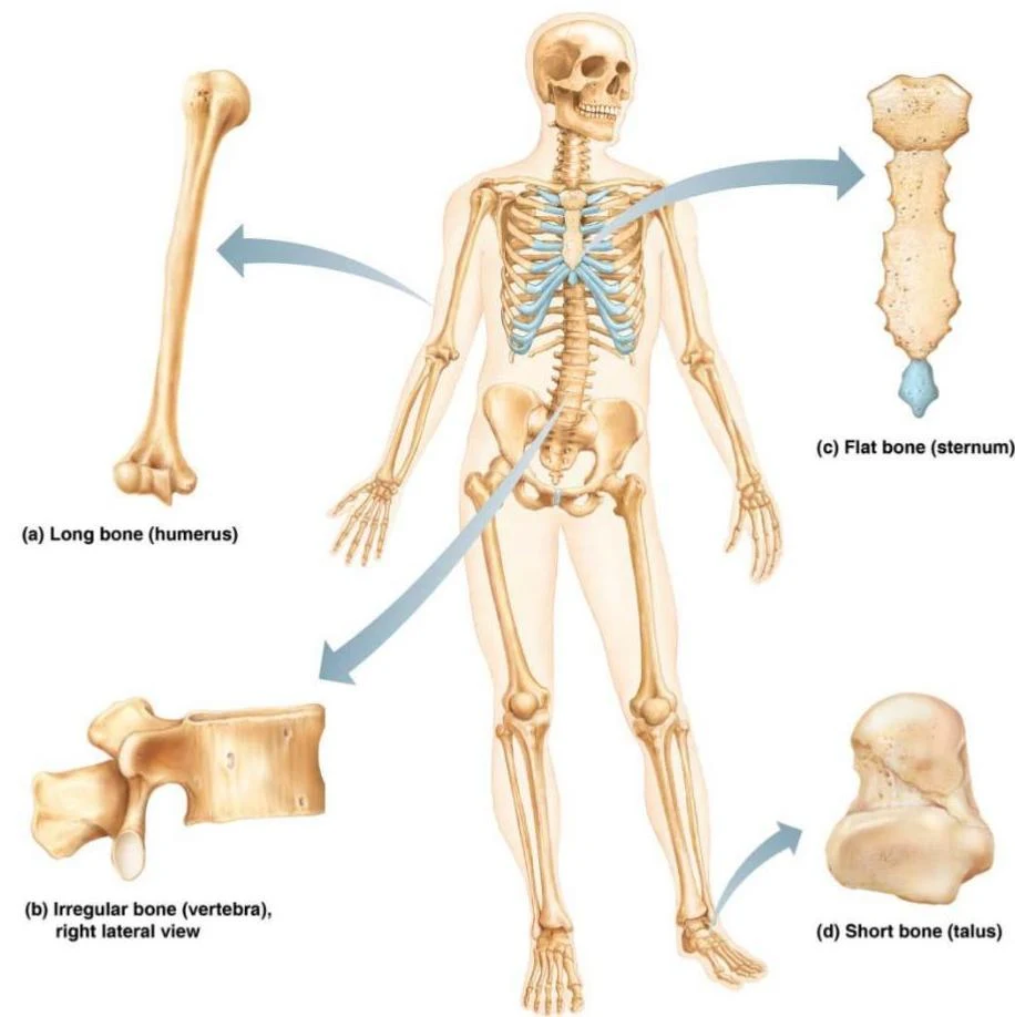
Long bones: These are longer than they are wide and typically function as levers for movement (e.g., limb bones).
Short bones: These are cube-shaped and are primarily found in the wrists and ankles.
Flat bones: These are thin, flat, and slightly curved. Examples include the sternum, scapulae, ribs, and most skull bones.
Irregular bones: These possess complex, non-uniform shapes, such as the vertebrae and hip bones.
Special bones: This category includes sesamoid bones, which form within tendons (such as the patella), and Wormian (sutural) bones, which are extra bone fragments found within the sutures of the skull.
This illustration displays different types of bones in the human skeletal system: long bone (humerus), irregular bone (vertebra), flat bone (sternum), and short bone (talus).
Gross Anatomy of Bone Tissue
Regardless of their shape, all bones are composed of two primary histological types: compact bone and spongy bone.
Compact and Spongy Bone
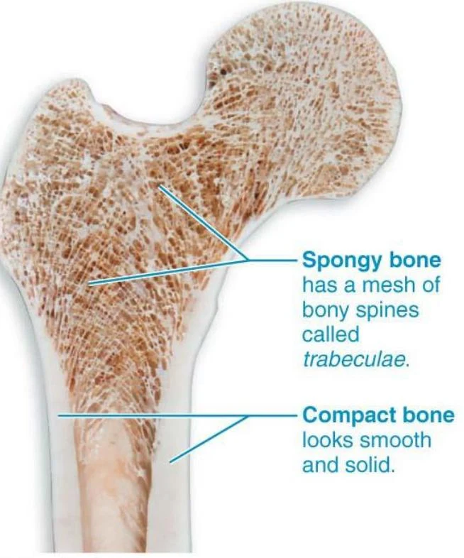
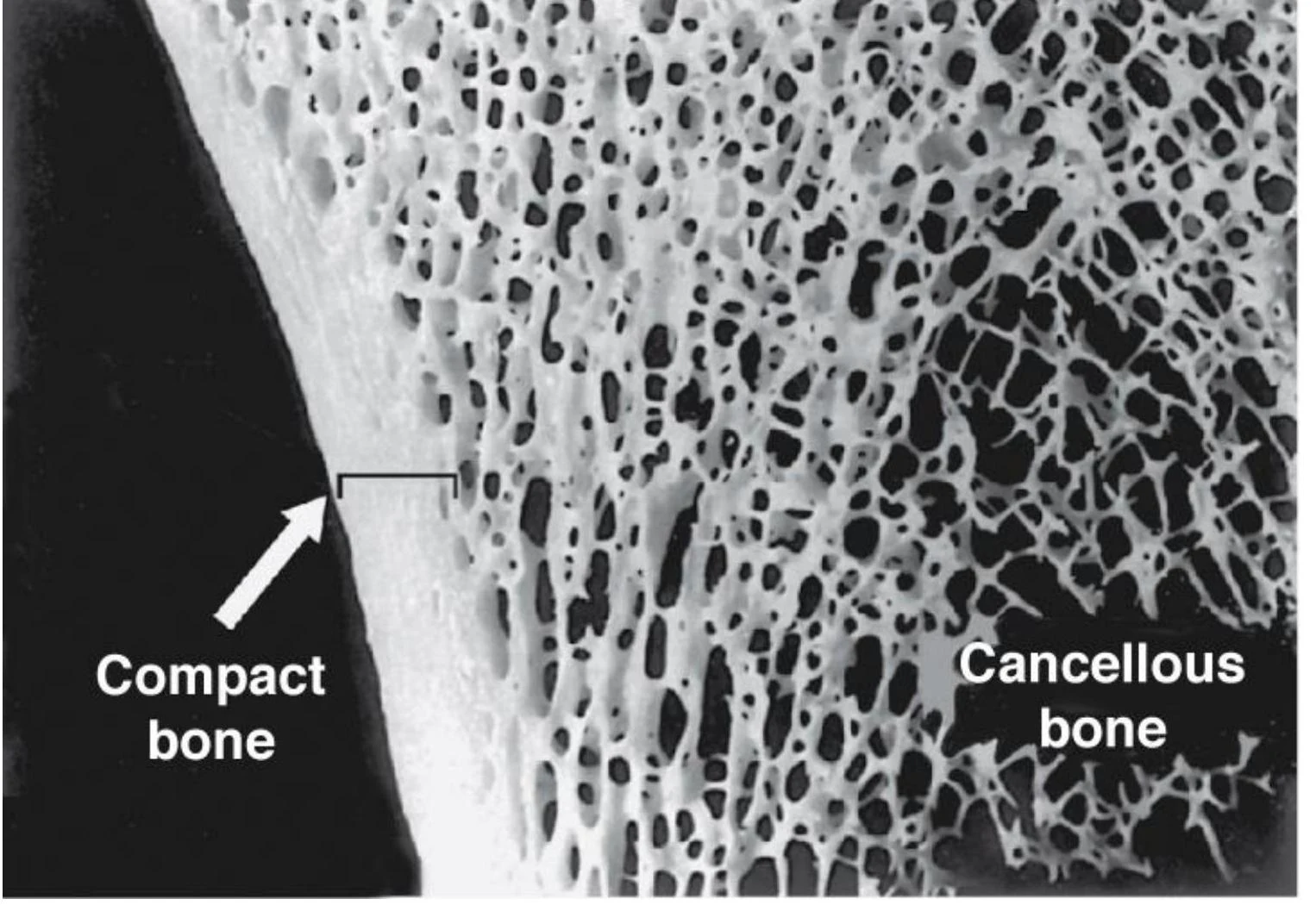
Compact bone is the dense, smooth outer layer that provides strength and resistance to compression. It makes up approximately 80% of all lamellar bone.
Spongy bone (also known as cancellous or trabecular bone) is located in the inner regions of bones. It consists of a honeycomb-like network of needle-like or flat pieces called trabeculae. These trabeculae are organized along lines of stress to help the bone resist mechanical pressure. The open spaces between these trabeculae are filled with bone marrow.
This illustration shows the internal structure of a bone, distinguishing between spongy bone, characterized by trabeculae, and compact bone, which appears smooth and solid.
Microscopic view of compact bone and cancellous bone structure.
In flat bones, the organization is unique: a layer of spongy bone, known as diploë, is sandwiched between two layers of compact bone.
Anatomy of a Typical Long Bone
A long bone is organized into several distinct regions:
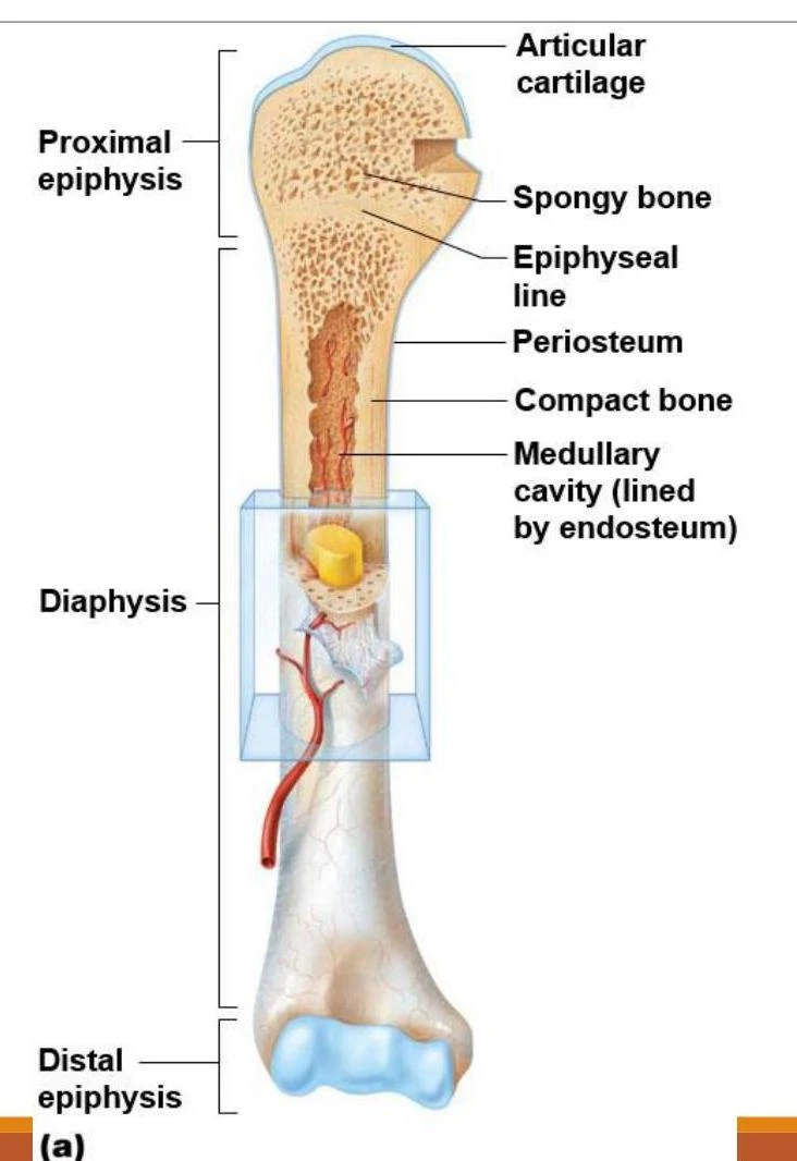
Diaphysis: The tubular shaft that forms the long axis of the bone. It consists of a thick layer of compact bone surrounding a central medullary cavity.
Epiphyses: The expanded ends of the bone. These consist of an external layer of compact bone and an internal core of spongy bone.
Epiphyseal line: The junction between the diaphysis and the epiphysis. This is the remnant of the childhood epiphyseal plate, which is the site of longitudinal bone growth.
Articular cartilage: A layer of hyaline cartilage that covers the joint surfaces to facilitate smooth movement.
Diagram illustrating the anatomy of a long bone, including the proximal epiphysis, diaphysis, and distal epiphysis, with labels for articular cartilage, spongy bone, epiphyseal line, periosteum, compact bone, and medullary cavity.
Bone Membranes
The bone is enclosed and protected by two distinct connective tissue membranes:
Periosteum: A white, double-layered membrane covering all external surfaces except for the joints.
The fibrous layer (outer) consists of dense irregular connective tissue and contains Sharpey’s fibers, which secure the membrane to the bone matrix.
The osteogenic layer (inner) contains primitive stem cells that give rise to bone cells. The periosteum also serves as an anchoring point for tendons and ligaments.
Endosteum: A delicate membrane that covers the internal bone surfaces, including the trabeculae of spongy bone and the lining of the canals passing through compact bone. Like the periosteum, it contains osteogenic cells.
Hematopoietic Tissue: Red vs. Yellow Marrow
The marrow found within bone cavities serves different physiological purposes:
Red Marrow (Hematopoietic tissue): This is responsible for hematopoiesis, the production of blood cells (red blood cells, white blood cells, and platelets). In newborns, red marrow is found in most bone cavities. In adults, it is primarily located in the trabecular cavities of spongy bone, the diploë of flat bones, and the heads of the femur and humerus.
Yellow Marrow: Found within the medullary cavity of adult long bones, this serves as an energy reserve. Notably, if a person becomes anemic, yellow marrow can convert back into red marrow to increase blood cell production.
Exam Tip: Remember that while newborns have red marrow throughout most of their skeleton, adults primarily restrict red marrow to the axial skeleton (flat bones) and the proximal ends of the humerus and femur.
Microscopic Anatomy and Bone Cells
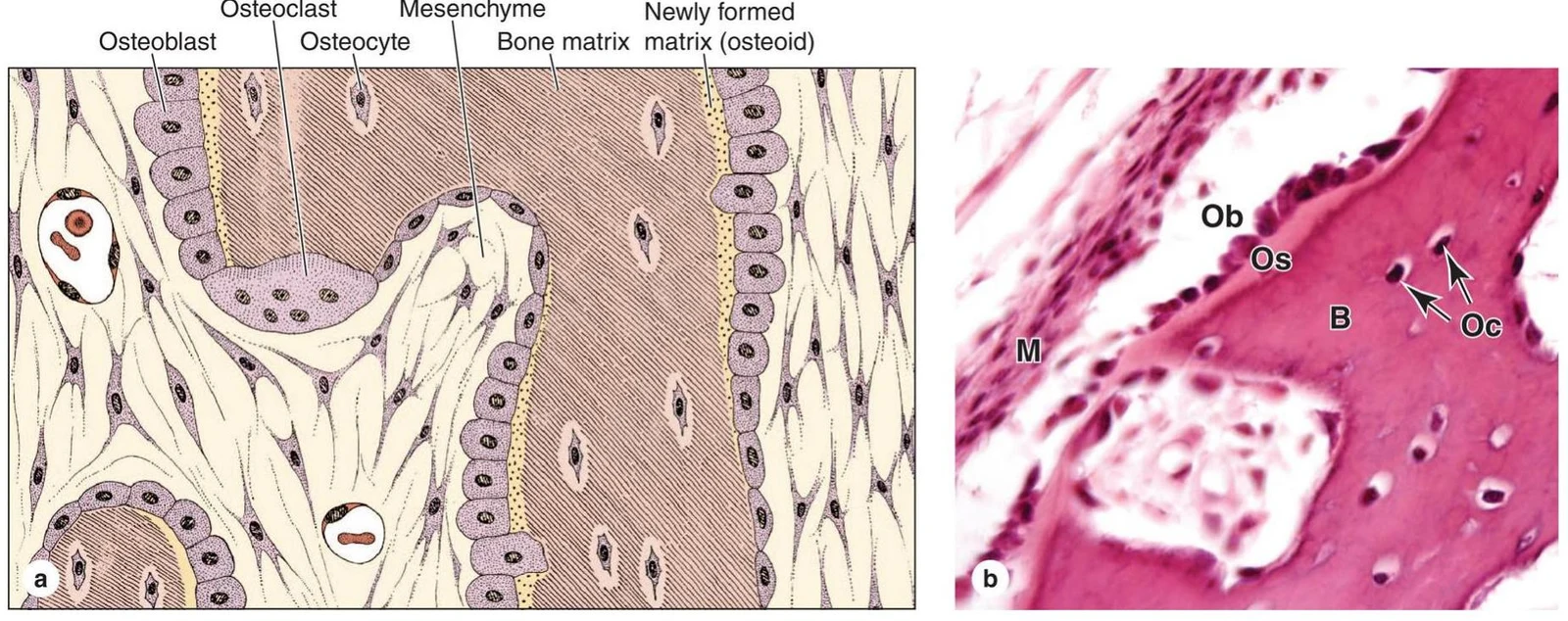
The bone matrix is composed of organic components (like collagen fibers for flexibility) and inorganic components (like calcium hydroxyapatite for hardness). This matrix is maintained and remodeled by four specialized cell types:
Osteogenic cells: Mitotically active stem cells found in the periosteum and endosteum that differentiate into osteoblasts.
Osteoblasts: Bone-forming cells that secrete osteoid (unmineralized bone matrix).
Osteocytes: Mature bone cells residing in small spaces called lacunae. They act as mechanical sensors, communicating with other cells to trigger remodeling in response to stress.
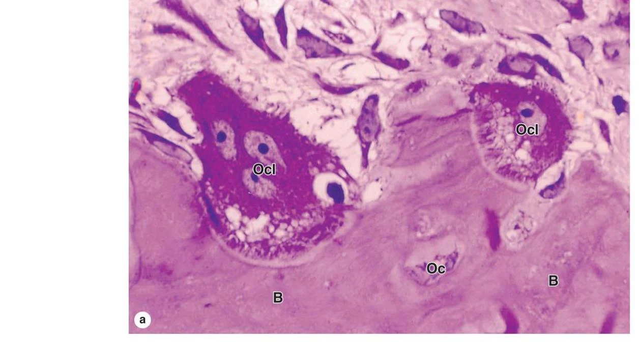
Osteoclasts: Large, multinucleated cells derived from the hematopoietic lineage. They function in bone resorption (the breakdown of bone) by using a ruffled border to secrete enzymes that degrade the matrix.
Diagrams illustrating bone formation: (a) A schematic representation of osteoblasts, osteocytes, osteoclasts, and bone matrix. (b) A micrograph showing bone tissue with osteoblasts (Ob), osteocytes (Oc) within lacunae, and bone matrix (B).
Histological image showing osteoclasts (Ocl) and osteocytes (Oc) within bone tissue (B).
Keep reading for free.
Create an account to finish this reading and make it part of your study routine.