Gastrointestinal Obstructions: Causes, Pathophysiology, and Clinical Presentation
This content explains gastrointestinal obstructions, detailing their causes, pathophysiology, and how their location influences clinical presentation and diagnosis. It covers esophageal, gastric outlet, and intestinal obstructions, including ileus and mechanical types like herniation, volvulus, adhesions, and intussusc
Gastrointestinal (GI) obstructions occur when the normal flow of contents through the digestive tract is impeded. These obstructions can be located anywhere from the esophagus to the large intestine, and their causes range from congenital defects to malignant tumors and mechanical issues like twisting or scarring. Understanding the pathophysiology of these obstructions is critical for clinical diagnosis, as the location of the blockage often dictates the patient's symptoms, such as the specific characteristics of their vomitus and the degree of abdominal distension.
Esophageal Obstruction and Dysphagia
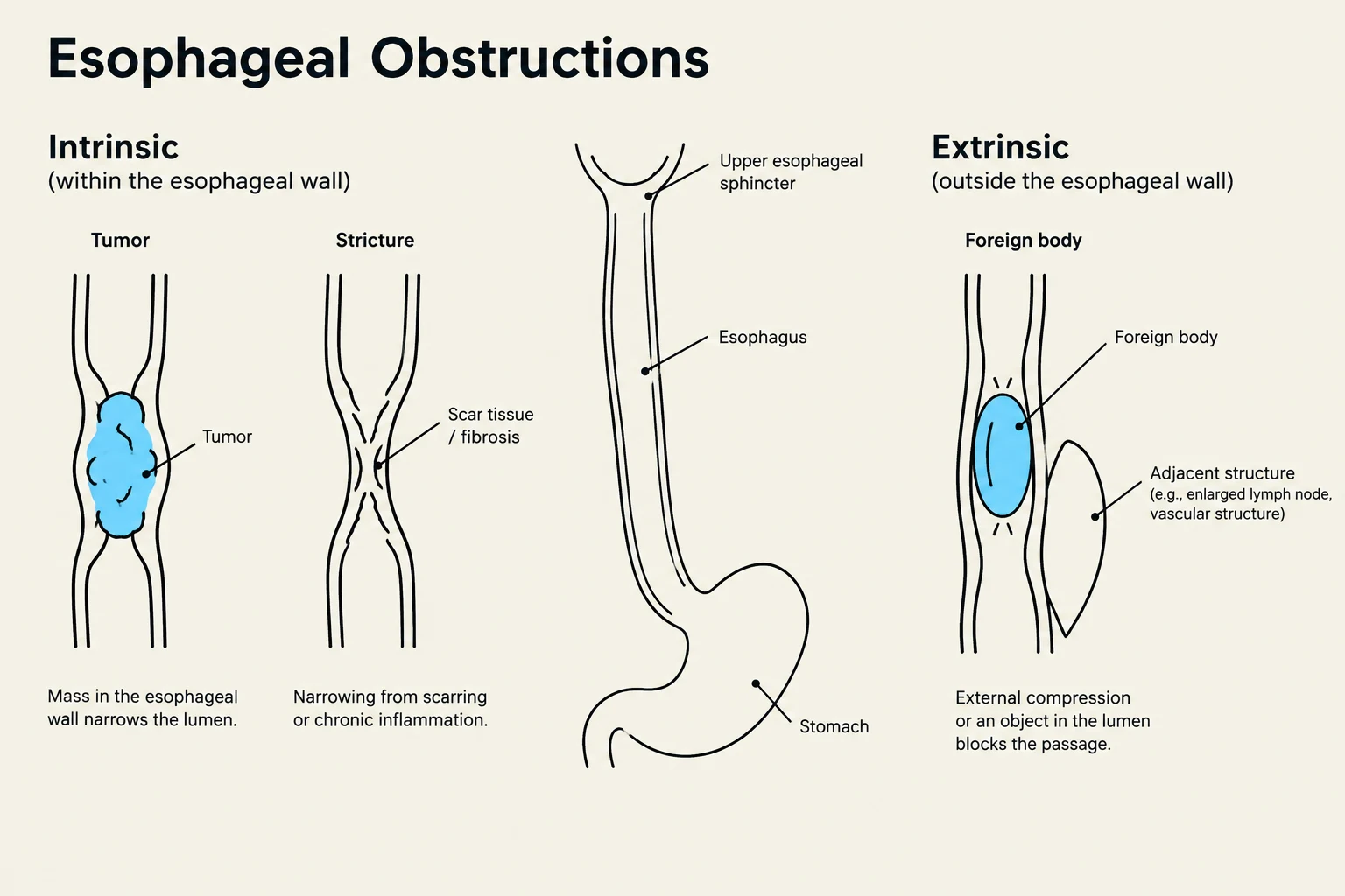
The earliest sign of an esophageal obstruction is often dysphagia, a medical term for difficulty swallowing. Dysphagia can stem from three primary categories:
Neurological/Autonomic issues: Damage to the nervous system (e.g., following a stroke) can compromise the voluntary or involuntary reflexes required for swallowing.
Intrinsic blockages: These originate within the esophageal wall, such as tumors, inflammation, or a narrowing known as a stricture.
Extrinsic blockages: These are caused by external objects, such as a swallowed foreign body that becomes lodged in the esophagus.
A unique clinical indicator for the location of an esophageal blockage is the timing of discomfort. If a patient feels pain 2 to 4 seconds after swallowing, the blockage is likely higher in the esophagus; if the discomfort is delayed by 10 to 15 seconds, the blockage is likely lower.
Another specific condition is achalasia, a rare disorder where the lower esophageal sphincter (cardiac sphincter) fails to relax. This causes food to sit in the lower esophagus, irritating the mucosal lining.
Clinical Alert: Aspiration Risk
One of the most significant complications of dysphagia is aspiration pneumonia. When a patient cannot effectively move saliva or food into the stomach, these substances may be inhaled into the trachea, leading to lung infection.
Diagrams illustrating intrinsic and extrinsic causes of esophageal obstructions, including tumors, strictures, foreign bodies, and external compression.
Esophageal Cancer Risk Factors
Esophageal cancer is a severe form of obstruction that is often asymptomatic until it reaches an advanced stage. There are two primary types, distinguished by their location and cellular origin:
Squamous Cell Carcinoma
This type typically affects the upper two-thirds of the esophagus. Key risk factors include:
Male sex at birth and age over 50.
History of smoking and alcohol consumption.
Consumption of betel nuts (common in Southeast Asia), which contain carcinogens.
Consumption of hot drinks or caustic substances.
Adenocarcinoma
This type affects the lower third of the esophagus. It is closely linked to Gastroesophageal Reflux Disease (GERD). Chronic exposure to gastric acid causes the esophageal cells to undergo a transformation into intestinal-like cells, a condition known as Barrett's esophagus. Individuals with Barrett's esophagus are at a significantly higher risk for adenocarcinoma.
Gastric Outlet Obstruction
Obstructions can also occur at the junction between the stomach and the duodenum, known as the gastric outlet. This area is controlled by the pyloric sphincter.
Causes: Obstructions here can be caused by tumors, inflammation from ulcers, or foreign bodies that have passed the esophagus but become lodged at the pyloric opening.
Congenital Pyloric Stenosis: This is a condition present at birth where the pyloric sphincter is abnormally narrow, preventing the passage of food and fluids. In infants, this often presents as difficulty feeding, weight loss issues, or frequent vomiting.
Intestinal Obstructions: Ileus and Mechanical Obstruction
When the obstruction moves into the intestines, it is categorized into two main types: ileus and mechanical obstruction.
Ileus
Ileus is a functional rather than a physical blockage. It describes a state where the ileum becomes "paralyzed" and fails to undergo peristalsis (the rhythmic contractions that move food). This is frequently seen after abdominal surgery due to the "over-manipulation" of the bowels or as a side effect of certain medications.
Mechanical Obstruction
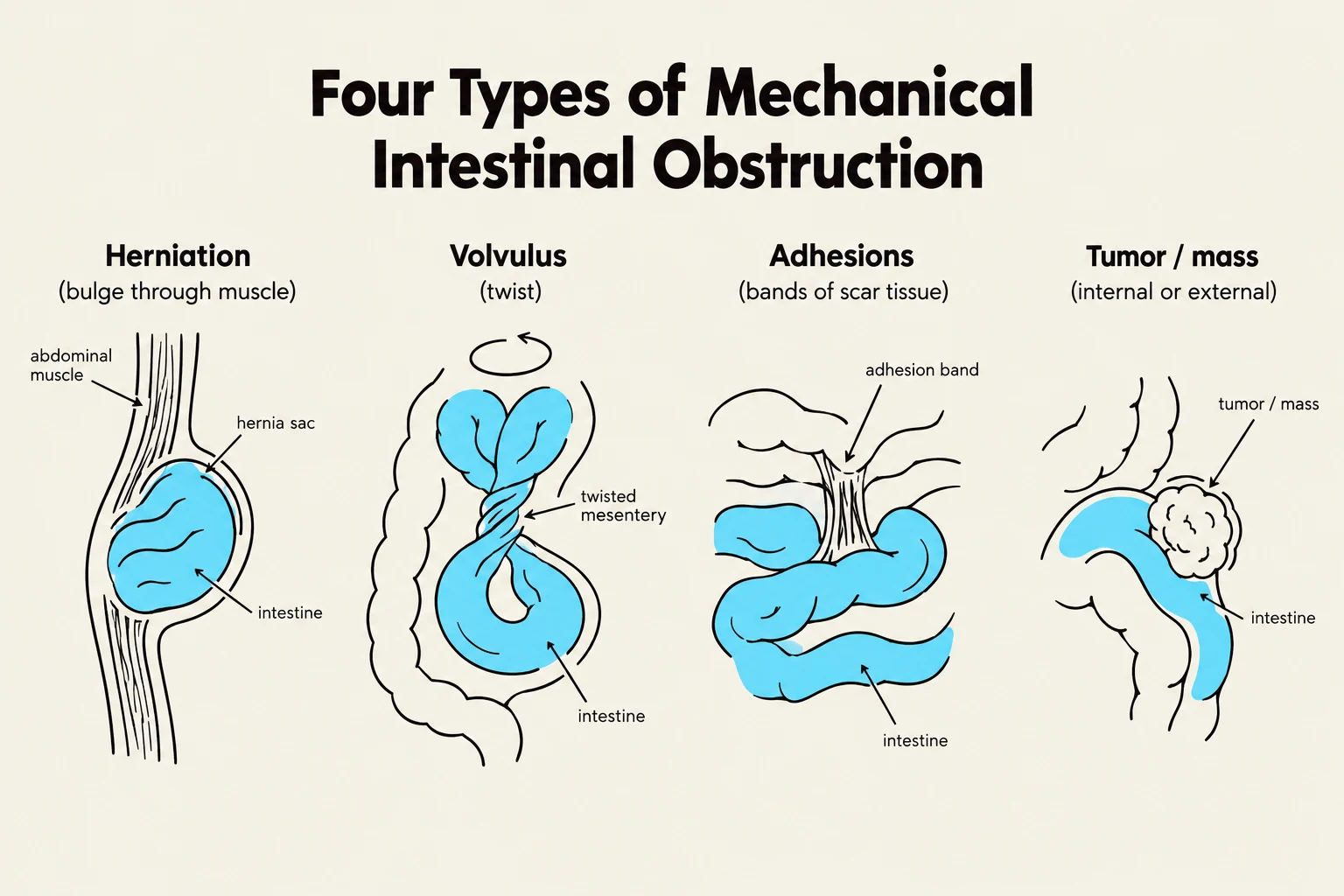
Mechanical obstruction involves a physical impediment to the movement of intestinal contents. There are four major subtypes:
Herniation: A portion of the abdominal muscle weakens, allowing a loop of the intestine to slip through and create a bulge. If the muscle wall pinches (strangulates) this loop, it can cut off blood supply and cause the tissue to die.
Volvulus: This occurs when a loop of the bowel physically twists upon itself. Like herniation, this carries a high risk of strangulation and bowel death.
Adhesions: These are bands of scar tissue that form between segments of the bowel, often following previous abdominal surgeries. These bands can prevent the bowel from moving adequately and are often very painful.
Intussusception: Often seen in pediatric patients, this is a "telescoping" effect where one piece of the bowel slips into another. This can also lead to strangulation and loss of blood supply.
Diagram illustrating four types of mechanical intestinal obstruction: herniation, volvulus, adhesions, and tumor/mass.
Clinical Presentation and Diagnosis
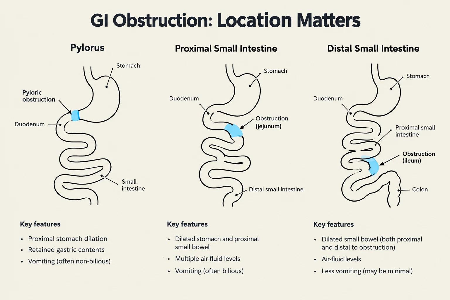
Clinicians use several indicators to locate an obstruction, most notably the characteristics of the patient's vomitus and the presence of abdominal distension.
Obstruction Location
Vomitus Characteristics
Abdominal Distension
Pylorus (Stomach/Duodenum junction)
Clear gastric fluid
Minimal/None
Proximal Small Intestine (Duodenum/Early Jejunum)
Bile-stained fluid
Mild
Distal Small Intestine
May be fecal in nature (if severe/chronic)
Significant
Exam Tip: The Bile Rule
If a patient is vomiting bile-stained fluid, the obstruction must be located below the point where the common bile duct empties into the duodenum.
To confirm a diagnosis, medical professionals utilize imaging such as X-rays, MRIs, or CT scans, as well as endoscopic or colonoscopic procedures to visualize the blockage directly. In some cases, exploratory surgery is required to identify and resolve the obstruction.
Diagrams illustrating the location of gastrointestinal obstructions in the pylorus, proximal small intestine, and distal small intestine, along with their key features.
Keep reading for free.
Create an account to finish this reading and make it part of your study routine.