This guide explains the fundamental physiological process of gas exchange, including ventilation, transport, and perfusion, and discusses clinical manifestations of impaired gas exchange, methods of assessment like auscultation, and strategies for prevention and management of respiratory conditions.
Gas exchange is the fundamental physiological process by which oxygen is transported to the body's cells and carbon dioxide is removed from them. This process relies on a delicate balance of ventilation (the movement of air in and out of the lungs), transport (the ability of hemoglobin to carry gases), and perfusion (the movement of blood through the pulmonary capillaries). When any part of this system fails, the body enters a state of respiratory distress that can quickly escalate from mild fatigue to life-threatening complications.
Recognizing Impaired Gas Exchange
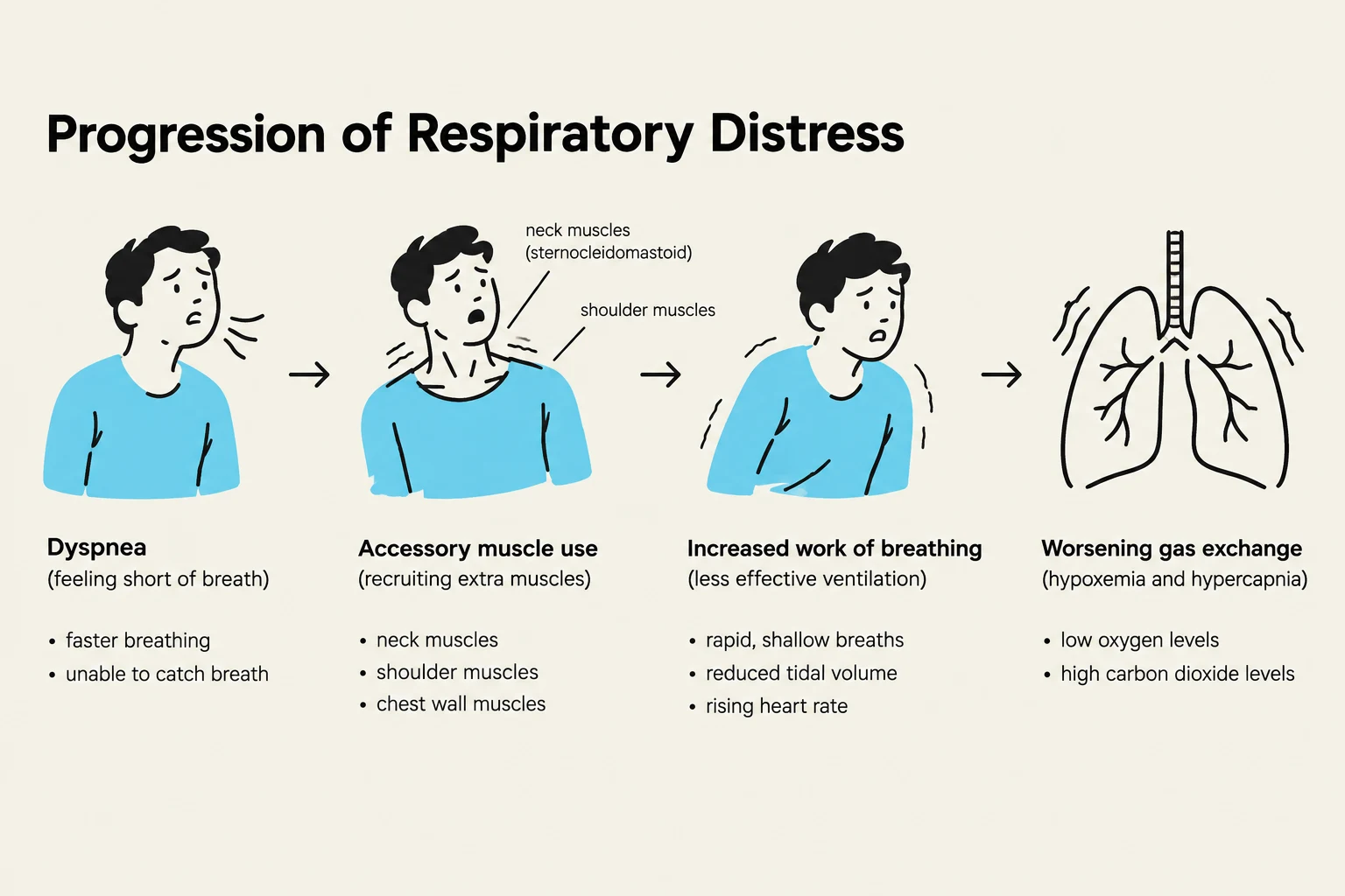
When gas exchange is inadequate, the body exhibits several clinical manifestations. Initially, a patient may experience dyspnea (difficult or painful breathing), anxiety, and the use of accessory muscles to assist with the work of breathing. As the condition progresses, the patient may develop hypoxemia, a state where there is insufficient oxygen reaching the cells or a drop in blood oxygen levels.
A critical late sign of severe hypoxemia is cyanosis, characterized by a bluish discoloration of the skin. In chronic conditions such as COPD (Chronic Obstructive Pulmonary Disease) or cystic fibrosis, the lungs may undergo structural changes, such as the development of a barrel chest due to air trapping. Furthermore, physical deformities like scoliosis (an abnormal curvature of the spine) can complicate respiratory function by causing asymmetric expansion of the thorax.
Clinical Alert: Pediatric Assessment
In infants and young children, respiratory distress may present differently than in adults. Watch for "grunting" on inspiration, nasal flaring, or the need to pause feeding to catch their breath.
This diagram illustrates the progression of respiratory distress, showing stages from dyspnea to accessory muscle use, increased work of breathing, and worsening gas exchange.
Clinical Assessment: Auscultating Lung Sounds
Auscultation is a vital skill for identifying the underlying cause of respiratory distress. While normal breath sounds include soft vesicular sounds heard over most of the lungs and louder bronchial sounds over the large airways, abnormal sounds provide clues to specific pathologies:
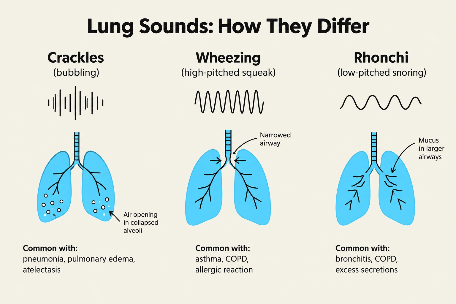
Crackles: These are popping, bubbling, or crackling sounds often caused by fluid in the airways, such as in heart failure, pulmonary edema, or atelectasis (the collapse of small lung sections).
Wheezing: High-pitched, squeaking, or musical sounds, typically heard during expiration, which are common in asthma or COPD.
Rhonchi: Low-pitched, snoring, or gurgling sounds caused by secretions in the large airways (e.g., bronchitis or pneumonia). These often improve after the patient coughs.
Stridor: A loud, harsh, crowing sound heard during inspiration. This is a medical emergency indicating an upper airway obstruction, such as epiglottitis or a foreign body.
Pleural Friction Rub: A grating or creaking sound produced by the rubbing of inflamed pleural surfaces.
This illustration differentiates between three types of lung sounds: crackles, wheezing, and rhonchi, detailing their associated waveforms, visual representations within the lungs, and common causes.
Understanding Asthma
Asthma is a specific chronic disorder characterized by periods of reversible airflow obstruction. It is driven by hyperreactive airways that respond to various triggers, causing the muscles surrounding the airways to contract and the tissues to become inflamed.
Common triggers include:
Genetics and family history.
Airborne allergens (pollen, dust, pet dander).
Emotional stress or exercise.
Respiratory infections.
Prevention and Management Strategies
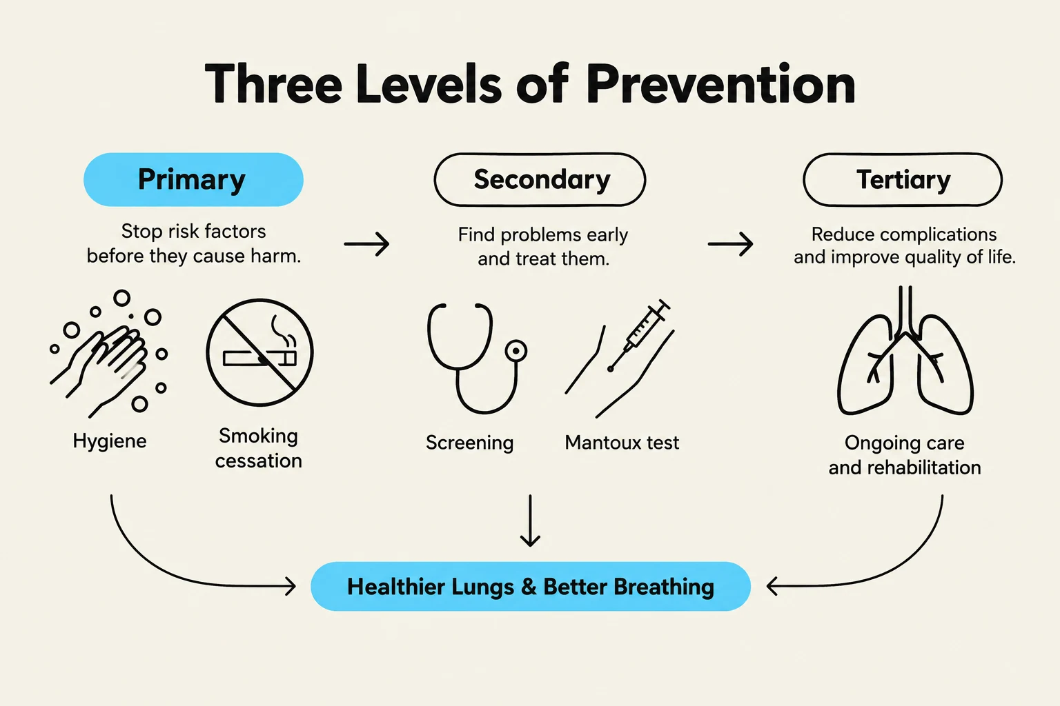
Effective respiratory care is organized into three levels of prevention: primary, secondary, and tertiary.
Primary Prevention: Health Promotion
Primary prevention aims to prevent the onset of disease through lifestyle and environmental controls:
Infection Control: Practicing diligent hand hygiene, cleaning frequently touched surfaces, and using respiratory etiquette (coughing into an elbow).
Smoking Cessation: Reducing or eliminating tobacco use to protect lung tissue.
Immunizations: Protecting against respiratory pathogens.
Postoperative Care: To prevent postoperative pulmonary complications like pneumonia or atelectasis, patients are encouraged to use an incentive spirometer to promote deep breathing and to cough regularly.
Venous Stasis Prevention: For immobile patients, preventing blood clots is essential. This is achieved through anticoagulants, elastic stockings, and early ambulation.
Secondary Prevention: Early Detection
Secondary prevention focuses on early diagnosis to shorten the duration and severity of a disease. An example is the administration of the Mantoux test to screen individuals for tuberculosis exposure.
Tertiary Prevention: Management of Established Disease
Tertiary prevention involves collaborative interventions to manage chronic conditions and prevent further deterioration:
Oxygen Therapy: Administering supplemental oxygen via nasal cannula, simple masks, or high-flow systems.
Pharmacological Treatments: Using bronchodilators to open airways.
Airway Management: Utilizing chest physiotherapy, suctioning, or postural drainage to clear secretions.
This diagram illustrates the three levels of prevention: primary (stopping risk factors), secondary (early detection and treatment), and tertiary (managing complications and improving quality of life), all contributing to healthier lungs and better breathing.
Exam Tip: Oxygen Safety
When managing oxygen therapy, always remember the fire safety rules: keep oxygen away from heat, flames, and smoking materials. Additionally, monitor for complications such as dry mucous membranes or carbon dioxide retention in chronic patients.
Keep reading for free.
Create an account to finish this reading and make it part of your study routine.