Explores the physiological processes of gas exchange in the respiratory system, including diffusion principles, oxygen and carbon dioxide transport via hemoglobin, and the regulation of breathing through chemoreceptors and brainstem centers. It also touches upon clinical imbalances like respiratory acidosis and alkalos
The primary goal of the respiratory system is to provide oxygen (O2) to the tissues for cellular metabolism and to remove carbon dioxide (CO2), a metabolic waste product. This exchange is not merely a matter of breathing air in and out; it is a sophisticated physiological process involving the movement of solutes across specialized membranes, the chemical transport of gases via blood proteins, and a tightly controlled feedback loop that adjusts breathing patterns to maintain acid-base homeostasis.
Principles of Gas Exchange
Gas exchange is driven by the process of diffusion, where molecules move from an area of higher partial pressure to an area of lower partial pressure. In the lungs, this occurs across the respiratory membrane (also known as the air-blood barrier). This membrane is an incredibly thin ultrastructure consisting of alveolar type I cells, the capillary endothelial cells, and a shared extracellular matrix.
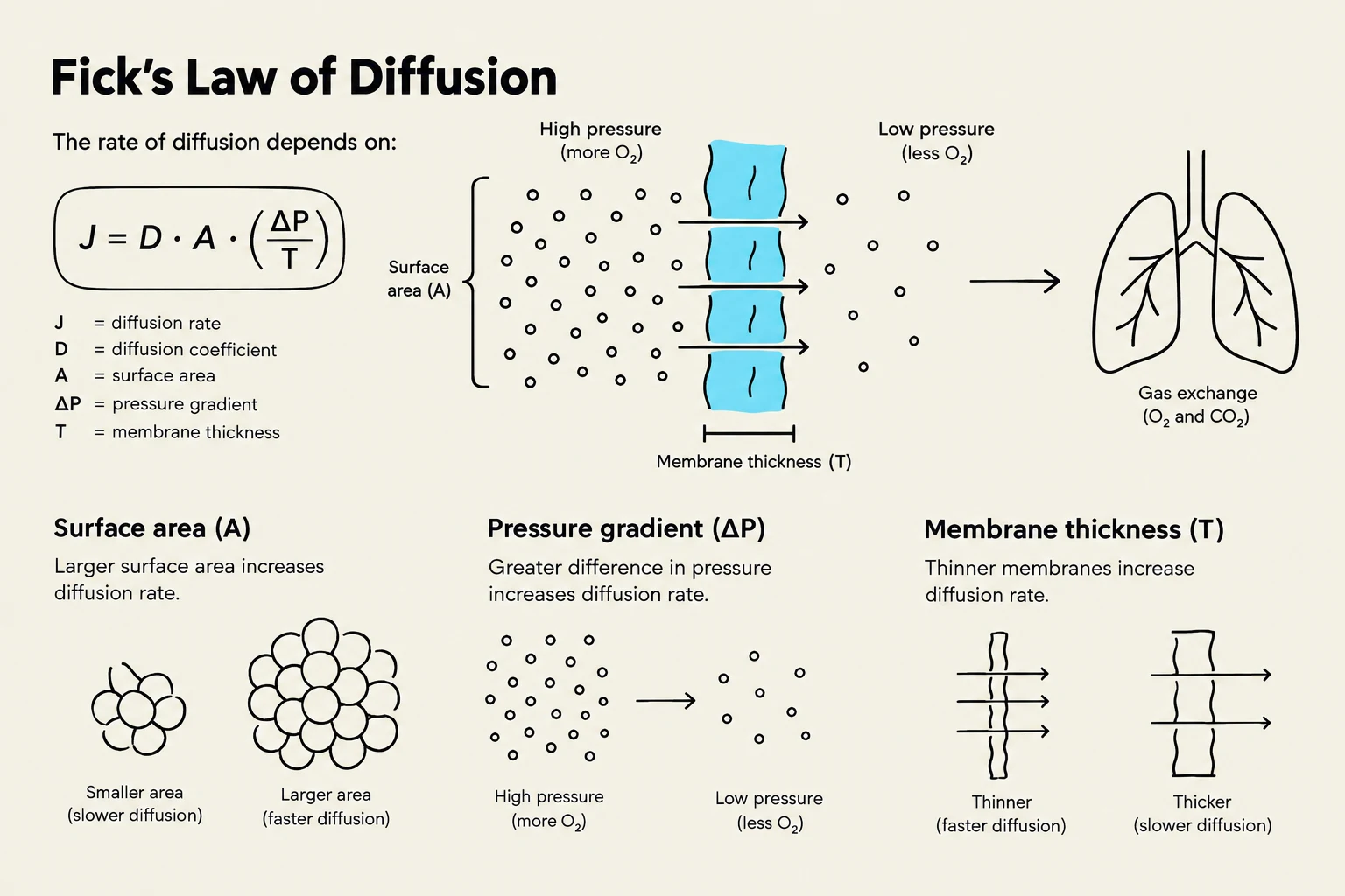
The efficiency of this diffusion is governed by Fick’s Law of Diffusion. The rate of gas transfer (Vgas) is determined by the following relationship:
VO2=A⋅D⋅dP1−P2
In this equation, A represents the available surface area, D is the diffusion constant (specific to the gas and the medium), P1−P2 is the partial pressure gradient across the membrane, and d is the diffusion distance. To maximize exchange, the body maintains a massive surface area (A) and a minimal membrane thickness (d).
Exam Tip: If a disease like pulmonary edema increases the fluid in the interstitial space, it increases the diffusion distance (d). According to Fick's Law, as d increases, the rate of diffusion (VO2) decreases, leading to hypoxia.
For gas exchange to be effective, the body must achieve ventilation-perfusion matching. This means that the amount of air reaching the alveoli (ventilation) must match the amount of blood reaching the capillaries (perfusion). The lungs utilize local autoregulation to optimize this: if an area of the lung has low alveolar O2, the local blood vessels undergo vasoconstriction. This shunts blood away from poorly ventilated areas toward well-ventilated alveoli, ensuring efficient oxygenation.
This diagram illustrates Fick's Law of Diffusion, explaining how the rate of diffusion is influenced by surface area, pressure gradient, and membrane thickness, with an application to gas exchange in the lungs.
Oxygen Transport and the Hemoglobin Curve
Once oxygen enters the blood, it must be transported to the tissues. Only about $1.5%$ of O2 is dissolved directly in the plasma; the remaining $98.5%$ is carried by hemoglobin (Hb) within red blood cells.
Hemoglobin is a highly efficient transporter due to a phenomenon called cooperativity. When the first O2 molecule binds to one of the four heme groups, it induces a conformational change in the protein, shifting it from a "Tense" (T-state) to a "Relaxed" (R-state). This shift increases the affinity of the remaining heme groups for oxygen, making subsequent binding much easier. This results in the characteristic sigmoidal (S-shaped) oxygen-hemoglobin dissociation curve.
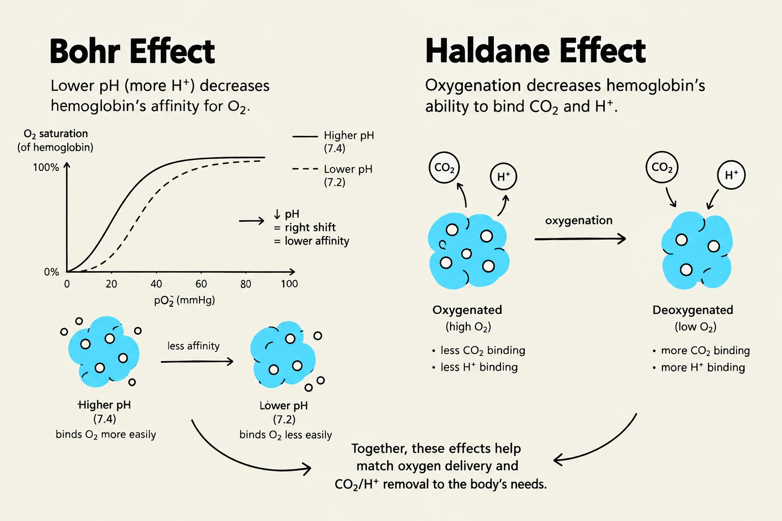
The ease with which hemoglobin releases oxygen to the tissues is influenced by the local chemical environment, a phenomenon known as the Bohr Effect. When tissues are metabolically active, they produce more CO2, $H^+$ (decreasing pH), and heat. These factors shift the dissociation curve to the right, meaning hemoglobin's affinity for O2 decreases, facilitating the unloading of oxygen where it is needed most.
Carbon Dioxide Transport and the Haldane Effect
Carbon dioxide transport is more complex than oxygen transport, involving three distinct mechanisms:
Dissolved in plasma: Approximately $7%$.
Carbaminohemoglobin: Approximately $23%$ of CO2 binds directly to the amino groups of hemoglobin.
Bicarbonate (HCO3−): The majority, about $70%$, is transported as bicarbonate.
The conversion of CO2 to bicarbonate is catalyzed by the enzyme carbonic anhydrase within red blood cells. The reaction follows this equilibrium:
CO2+H2O↔HCO3−+H+
As bicarbonate is produced, it is often moved out of the red blood cell into the plasma in exchange for a chloride ion to maintain electrical neutrality; this is known as the chloride shift.
Complementing this is the Haldane Effect, which describes how the oxygenation of blood affects CO2 transport. In the lungs, when O2 binds to hemoglobin, the hemoglobin becomes more acidic, which promotes the release of CO2 from the blood into the alveoli for exhalation.
The Bohr and Haldane effects illustrate how hemoglobin's affinity for oxygen, carbon dioxide, and hydrogen ions changes with pH and oxygen levels, facilitating efficient gas exchange in the body.
Regulation of Respiration
The body regulates breathing through a negative feedback loop designed to maintain stable levels of PCO2, PO2, and pH.
Sensors and Integrating Centers
The system relies on two types of chemoreceptors:
Central chemoreceptors: Located in the medulla oblongata, these are highly sensitive to changes in the pH of the brain's extracellular fluid, which is directly influenced by arterial CO2 levels.
Peripheral chemoreceptors: Located in the carotid and aortic bodies, these monitor arterial O2, CO2, and pH.
These sensors send signals to the integrating centers in the brainstem. The medulla oblongata contains the primary respiratory groups (the Ventral Respiratory Group, VRG, and Dorsal Respiratory Group, DRG) that act as the pacemaker for rhythm. The pons contains centers that modulate this rhythm to ensure smooth transitions between inhalation and exhalation.
Clinical Imbalances
When the regulatory loop fails, several clinical conditions can arise:
Hypoventilation: Reduced breathing rate or depth leads to an accumulation of CO2 (hypercapnia). This increases $H^+$ concentration, resulting in respiratory acidosis.
Hyperventilation: Excessive breathing lowers arterial PCO2. This reduction in CO2 shifts the equilibrium to consume $H^+$, leading to respiratory alkalosis and potentially causing cerebral vasoconstriction.
Clinical Note: Hypoxic Drive
In patients with chronic hypercapnia (persistently high CO2), the central chemoreceptors may become desensitized. These patients may rely on their "hypoxic drive"—the peripheral chemoreceptors' response to low O2—to stimulate breathing. In such cases, administering high concentrations of supplemental O2 can inadvertently suppress their drive to breathe, leading to dangerous hypoventilation.
This diagram illustrates the negative feedback loop that regulates breathing rate based on blood gas levels.
Keep reading for free.
Create an account to finish this reading and make it part of your study routine.