GABAergic and Glutamatergic Neurotransmission in the CNS
Explores the balance between excitatory (Glutamate) and inhibitory (GABA) neurotransmission in the central nervous system, detailing their mechanisms, receptors, clinical significance, and pharmacology.
The human brain contains approximately 100 billion neurons that work in concert to manage everything from movement and sensation to complex cognition and emotion. To maintain stability within these neural networks, the nervous system relies on a precise balance between excitation and inhibition. This balance is primarily governed by two key neurotransmitters: Glutamate, the brain's primary excitatory signal, and γ-aminobutyric acid (GABA), its primary inhibitory signal.
The Nervous System Architecture
The nervous system is broadly divided into two main components:
Central Nervous System (CNS): Comprising the brain and the spinal cord.
Peripheral Nervous System (PNS): The network of nerves that branch off from the spinal cord to reach all parts of the body.
Within these systems, neurons communicate through two distinct methods: electrical transmission, which utilizes gap junctions for rapid, direct signaling, and chemical transmission, which relies on the release of neurotransmitters across a synapse.
Diagrams illustrating the mechanisms of chemical and electrical synapses in neurons.
The Mechanics of Neurotransmission
At the cellular level, communication is driven by the action potential, a rapid change in the electrical potential of a neuron's membrane. When a neuron receives a signal, it undergoes changes in ion conductance—the movement of ions like Na+, K+, Ca2+, or Cl− across the membrane.
Excitatory Neurotransmission (The "Go" Signal)
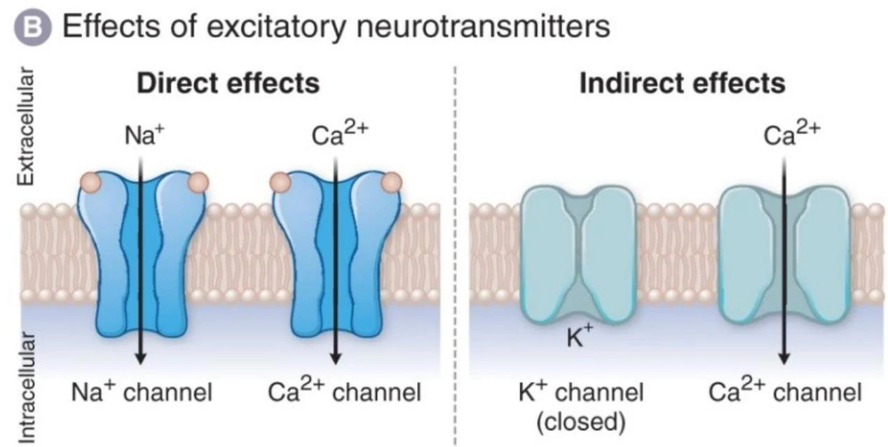
Glutamate is the major excitatory neurotransmitter. It functions by inducing depolarization, a process where the membrane potential becomes more positive, moving it closer to the threshold required to trigger an action potential. This is typically achieved through the influx of cations, specifically Na+ and Ca2+. The resulting increase in the likelihood of neuronal firing is known as an Excitatory Postsynaptic Potential (EPSP).
Glutamate acts on two main types of receptors:
Ionotropic receptors: These include AMPA, Kainate, and NMDA receptors. These receptors act as ligand-gated ion channels that allow for rapid ion influx. The NMDA receptor is particularly notable because it allows Ca2+ influx, which is critical for learning and memory.
Metabotropic receptors: Known as mGluRs, these act through slower second messenger systems rather than direct ion channels.
Inhibitory Neurotransmission (The "Stop" Signal)
GABA is the primary inhibitory neurotransmitter, providing the "stop" signal to prevent excessive neuronal activity. It induces hyperpolarization, making the membrane potential more negative and moving it away from the firing threshold. This results in an Inhibitory Postsynaptic Potential (IPSP).
GABA achieves this through two mechanisms:
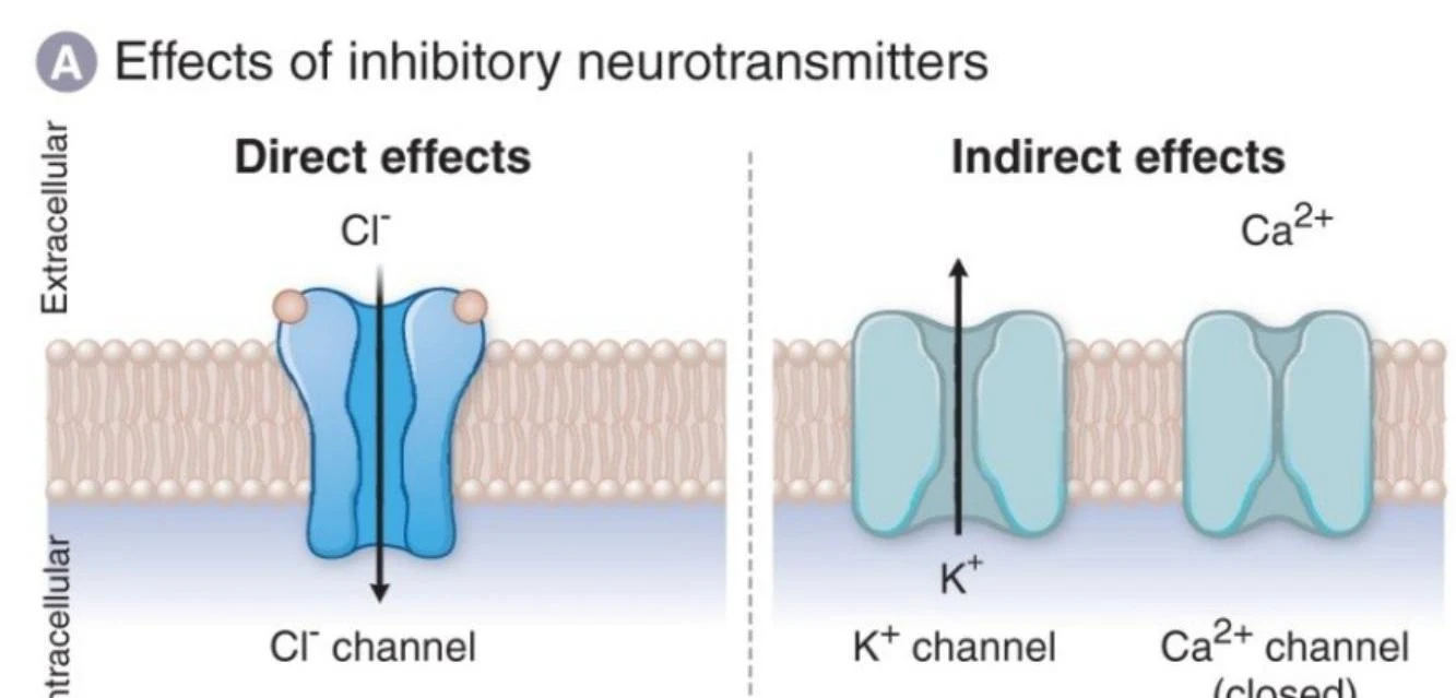
Direct effects: Binding to GABAA (ionotropic) receptors, which opens Cl− channels, leading to an influx of chloride ions.
Indirect effects: Binding to GABAB (metabotropic) receptors, which activates G-proteins to open K+ channels, leading to an efflux of potassium ions.
Diagram illustrating the direct and indirect effects of excitatory neurotransmitters on ion channels, specifically showing Na+, Ca2+, and K+ channels in the cell membrane.
Diagram illustrating the direct and indirect effects of inhibitory neurotransmitters on ion channels, specifically chloride (Cl-) and potassium (K+) channels, and calcium (Ca2+) channels.
Clinical Significance: When Balance is Lost
The delicate equilibrium between Glutamate and GABA is essential for stable neural networks. When this balance is disrupted, it often results from channelopathies—malfunctions in the Na+, K+, Ca2+, or Cl− channels.
Imbalance Type
Physiological Result
Clinical Manifestations
Reduced GABA Signaling
Increased excitability (Reduced IPSPs)
Anxiety, Seizures
Increased GABA Signaling
Excessive inhibition (Increased IPSPs)
Sedation, Motor incoordination, Respiratory depression, Coma
Exam Tip: If a question asks about the mechanism of neurodegeneration in the context of glutamate, look for "excitotoxicity" driven by excessive Ca2+ influx through NMDA receptors.
Pharmacology of the GABAA Receptor
The GABAA receptor is a pentameric (five-subunit) transmembrane protein that serves as a major target for various drugs. Because it has multiple allosteric binding sites, different classes of drugs can modulate its function in distinct ways.
Modulators of GABAA
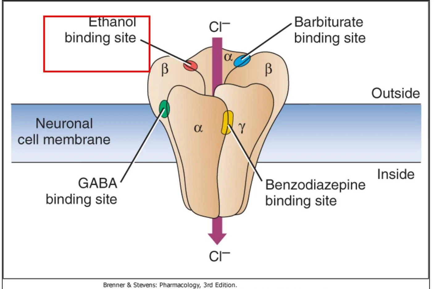
Benzodiazepines: These bind to an allosteric site between the α and γ subunits. They facilitate GABA binding, which increases the frequency of the chloride channel opening.
Barbiturates: These bind adjacent to the α and β subunits. They increase the duration of the chloride channel opening, even in the absence of GABA.
Ethanol: Alcohol binds to the receptor and enhances chloride influx, contributing to its sedative effects.
Diagram illustrating the GABA-gated chloride channel in a neuronal cell membrane, showing binding sites for ethanol, barbiturates, benzodiazepines, and GABA, and the movement of chloride ions.
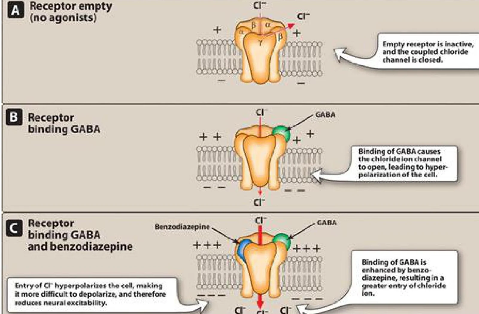
This diagram illustrates the mechanism of GABA and benzodiazepine action on a chloride ion channel receptor. Panel A shows an empty receptor that is inactive. Panel B depicts GABA binding to the receptor, causing the chloride ion channel to open and leading to hyperpolarization. Panel C shows that benzodiazepine enhances GABA binding, resulting in a greater influx of chloride ions and reduced neural excitability.
Neurotransmitter Metabolism and Recycling
The brain maintains these levels through complex metabolic cycles. For example, the enzyme Glutamic Acid Decarboxylase (GAD) converts glutamate into GABA using Vitamin B6 as a cofactor.
Furthermore, the glutamate-glutamine cycle involves a partnership between neurons and glial cells. Glial cells use the enzyme glutamine synthetase to transform excess glutamate into glutamine, which is then transported back to the neuron to be converted back into glutamate by glutaminase. This recycling ensures that the synaptic cleft is cleared of excitatory neurotransmitters, preventing excitotoxicity.
Keep reading for free.
Create an account to finish this reading and make it part of your study routine.