Fungal Pathogenesis and Diagnostic Methods in Microbiology
An educational overview of mycology, distinguishing between microbes and pathogens, explaining fungal pathogenesis mechanisms, and detailing diagnostic methods like isolation and molecular detection.
Understanding mycology is a cornerstone of veterinary and medical microbiology. Fungi are eukaryotic, chemo-organotrophic organisms that exhibit immense diversity, ranging from single-celled yeasts to complex, multicellular molds. Whether dealing with a superficial skin infection in a domestic pet or a life-threatening systemic infection in a large animal, a clinician must understand how these organisms interact with the host and how to accurately identify them in a laboratory setting.
Microbes vs. Pathogens: A Critical Distinction
Before diving into the complexities of fungal disease, it is essential to clarify fundamental terminology. In clinical microbiology, the terms "microbe" and "pathogen" are often used interchangeably, but they have distinct meanings:
Microbe: This is a broad, umbrella term encompassing all microscopic organisms, including bacteria, viruses, fungi, and protozoa.
Pathogen: This refers specifically to a microbe that has the capacity to cause disease.
While every pathogen is a microbe, not every microbe is a pathogen. Many microbes exist as commensals or saprophytes (living on decaying organic matter) without causing harm to a host.
The Mechanisms of Fungal Pathogenesis
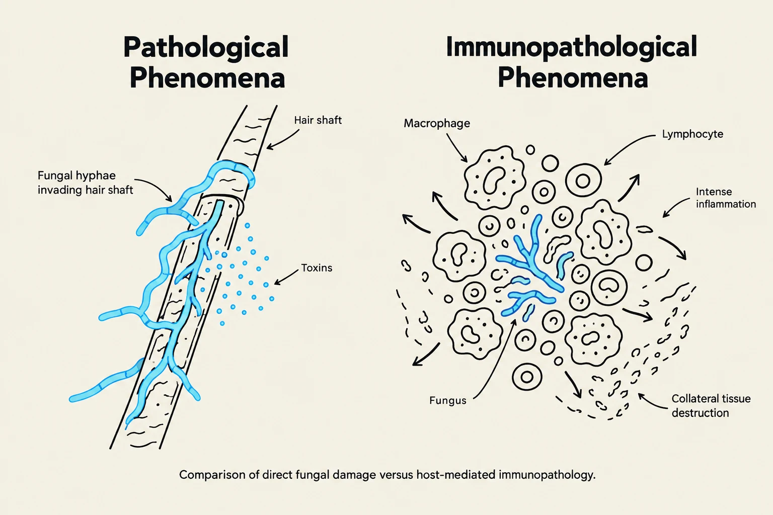
Comparison of direct fungal damage versus host-mediated immunopathology, illustrating fungal hyphae invading a hair shaft and the immune response involving macrophages and lymphocytes leading to inflammation and tissue destruction.
Fungal pathogenesis is defined by the interplay between the organism's virulence factors and the host's immune response. Infections can be categorized by their depth and spread: superficial/cutaneous mycoses affect the keratinized layers of the skin, hair, and nails, while deep or systemic mycoses impact internal organs and can lead to death.
Pathological and Immunopathological Phenomena
The damage caused by a fungus is not always a direct result of the organism "eating" tissue. Instead, damage often arises from two distinct phenomena:
Pathological Phenomena: This refers to the direct damage caused by the fungus, such as the invasion of hair shafts by dermatophytes or the production of toxins.
Immunopathological Phenomena: This occurs when the host's own immune system causes damage. If the body cannot effectively expel the fungus, it may trigger intense, chronic inflammation. This intense inflammatory response can lead to significant tissue destruction, often more severe than the damage caused by the fungus itself.
The Role of the Cell-Mediated Response
A critical factor in determining the progression of a fungal infection is the host's cell-mediated response. While innate immunity (such as neutrophils and macrophages) provides the first line of defense, a robust cell-mediated immune response is necessary to control deep mycoses. A weak or compromised cell-mediated response can allow a localized infection to become multifocal or progress into a life-threatening disseminated disease.
Clinical Note: Mixed Infections
Many systemic fungal diseases (like histoplasmosis) compromise the host's immune system. This often leads to "mixed infections," where a primary fungal pathogen allows for secondary bacterial or parasitic infections to take hold.
Fungal Disease Diagnosis: Isolation vs. Detection
When a clinician suspects a fungal infection, they must choose between two primary diagnostic pathways: isolation and detection. Understanding the difference is vital for selecting the correct test and managing patient expectations regarding turnaround time.
Isolation: The Gold Standard for Identification
Isolation involves culturing the organism from a clinical sample (such as a skin scraping, hair pluck, or nasal swab) onto specific growth media, such as Sabouraud Dextrose Agar (SDA).
Why it is prioritized: Fungi are highly complex. While some methods can tell you a fungus is present, isolation is often required for species-level identification. By growing the organism on a plate, microbiologists can observe macroscopic characteristics (color, texture, growth rate) and microscopic structures (spore types, hyphal arrangement) to confirm the exact species.
The Challenge: Fungi grow much slower than bacteria. While bacteria might grow overnight, dermatophytes may require 7 to 21 days to produce visible colonies.
Detection: Rapid Molecular Identification
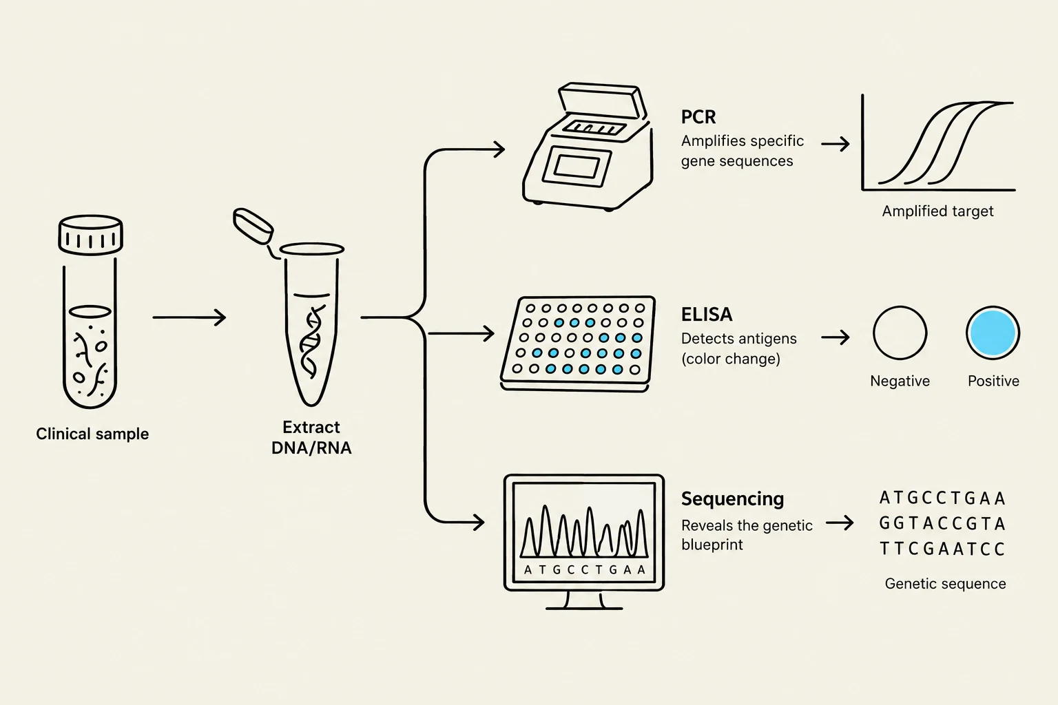
This diagram illustrates common molecular biology techniques used in genetic analysis, including PCR for gene amplification, ELISA for antigen detection, and DNA sequencing to reveal genetic blueprints. It shows the process from a clinical sample to extracted DNA/RNA, followed by different analytical methods and their typical outputs.
Detection focuses on identifying the presence of the pathogen's molecular components rather than growing the living organism itself. This is particularly useful when a rapid diagnosis is needed or when the organism is difficult to isolate from a specific sample (e.g., cerebrospinal fluid).
PCR (Polymerase Chain Reaction): This method targets specific DNA or RNA sequences. For successful species-specific identification, the test must target highly specific genes unique to that pathogen.
ELISA (Enzyme-Linked Immunosorbent Assay): An immunological method used to detect specific antigens or antibodies related to the fungal infection.
Sequencing: Used for the most precise identification, sequencing provides the genetic blueprint of the pathogen, allowing for definitive classification.
Summary Table: Diagnostic Comparison
Feature
Isolation (Culture)
Detection (Molecular/Immuno)
Primary Target
Living organism
DNA, RNA, or Antigens
Method
Growth on agar plates (e.g., SDA)
PCR, ELISA, Sequencing
Speed
Slow (up to 21 days)
Rapid
Strength
Species-level identification
Rapid presence/absence confirmation
Weakness
Slow growth; may fail if patient is on antifungals
Requires specific primers/antibodies
Exam Tip: Clinical Reasoning
If a question asks why isolation is preferred over simple detection in complex fungal cases, the answer is usually that isolation allows for definitive species-level identification through the observation of morphological structures.
Safety and Practical Considerations
Working with fungi carries inherent risks. Many molds produce highly infectious aerosols that can be inhaled by laboratory staff or clinicians.
Containment: Suspected pathogenic molds should never be opened outside of appropriate containment (like a biosafety cabinet).
Personal Protection: Always wear gloves and appropriate PPE, especially when handling skin lesions or suspected zoonotic samples (diseases that can jump from animals to humans), such as dermatophytes.
Sample Submission: Always contact the laboratory before sending samples, as certain pathogens (like endemic dimorphic fungi) require specialized handling and advanced containment protocols.
Keep reading for free.
Create an account to finish this reading and make it part of your study routine.