This content explores drug-receptor interactions, binding site specificity, and the mechanisms of drug action through receptors or nonreceptor-mediated processes.
Pharmacodynamics is defined as the study of what a drug does to the body. While pharmacokinetics focuses on the movement of a drug through the system (absorption, distribution, metabolism, and elimination), pharmacodynamics delves into the mechanism of action (MOA)—the detailed study of how a drug produces a specific pharmacologic effect. This process typically begins when a drug molecule interacts with a specific biological target, such as a receptor, enzyme, or structural protein, triggering a cascade of events that results in a therapeutic or toxic response.
Fundamentals of Drug-Receptor Interactions
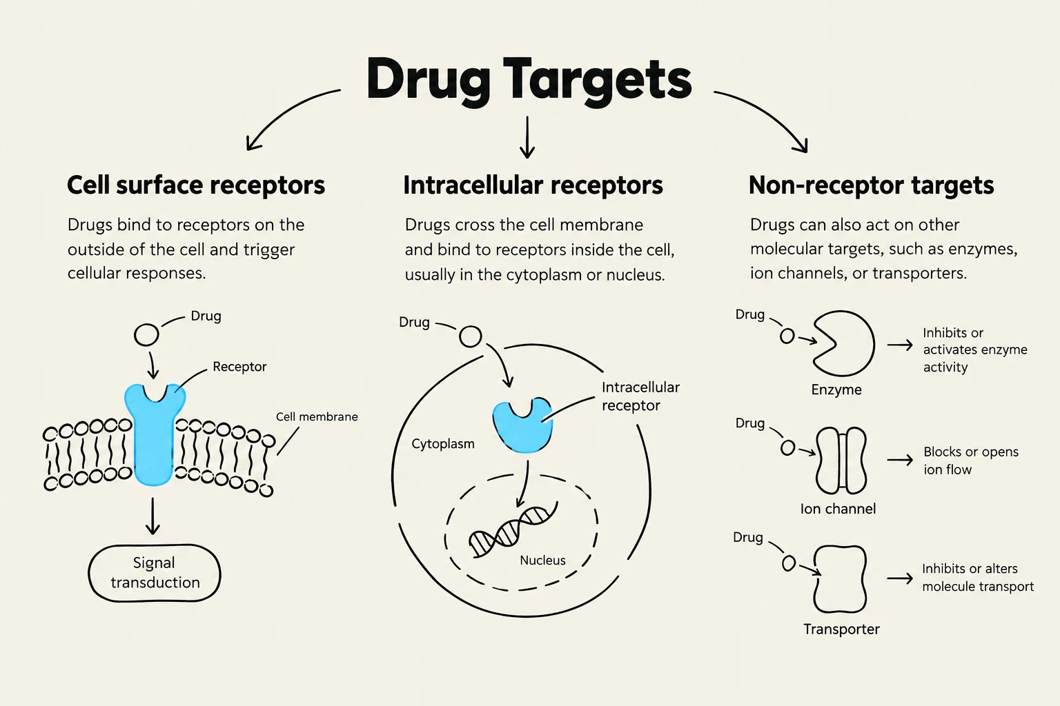
Most drugs exert their effects by binding to macromolecular components of a cell known as receptors or drug targets. These targets can be located on the cell surface, within intracellular compartments (like the nucleus or organelles), or even in the extracellular space, such as coagulation factors. When a drug binds to its target, it alters a cellular or physiological response.
The Drug-Receptor Occupancy Theory suggests that a response occurs when a sufficient number of receptors are occupied by a drug or an endogenous ligand. The cumulative effect of this occupancy becomes apparent at the cellular level, and when this occurs across many cells, it manifests as a visible response in an organ or the entire organism.
Binding Specificity and Forces
Not every drug can bind to every receptor. Binding site specificity is determined by the unique three-dimensional structure, shape, and electrical charge of both the drug and the binding site. The local chemical environment—including hydrophilicity, hydrophobicity, and pKa—also plays a critical role.
The interaction between a drug and its receptor is driven by intermolecular forces. These can be:
Attractive forces: These can be reversible (varying in strength) or irreversible (such as a covalent bond).
Repulsive forces: These prevent binding.
The "Induced Fit Theory" suggests that the binding process often involves conformational changes in the receptor to accommodate the drug, which is what ultimately initiates the biological response.
Exam Tip: Remember that "more receptor types bound" generally means a drug is "less specific," which often leads to more unwanted side effects.
Nonreceptor-Mediated Mechanisms
While most drugs use receptors, some work through nonreceptor-mediated mechanisms:
Chemical Actions: Antacids neutralize stomach acid through a direct chemical reaction (CaCO3+2HCl→CaCl2+H2O+CO2). Chelating agents, such as Calcium EDTA, bind to heavy metals like lead to remove them from the body.
This diagram illustrates the three main types of drug targets: cell surface receptors, intracellular receptors, and non-receptor targets, explaining how drugs interact with each to elicit a cellular response.
Receptor Transduction Mechanisms
Once a drug binds to a receptor, the process of converting that binding event into a cellular response is known as transduction. Receptors are classified based on their specific transduction components.
1. G Protein-Coupled Receptors (GPCRs)
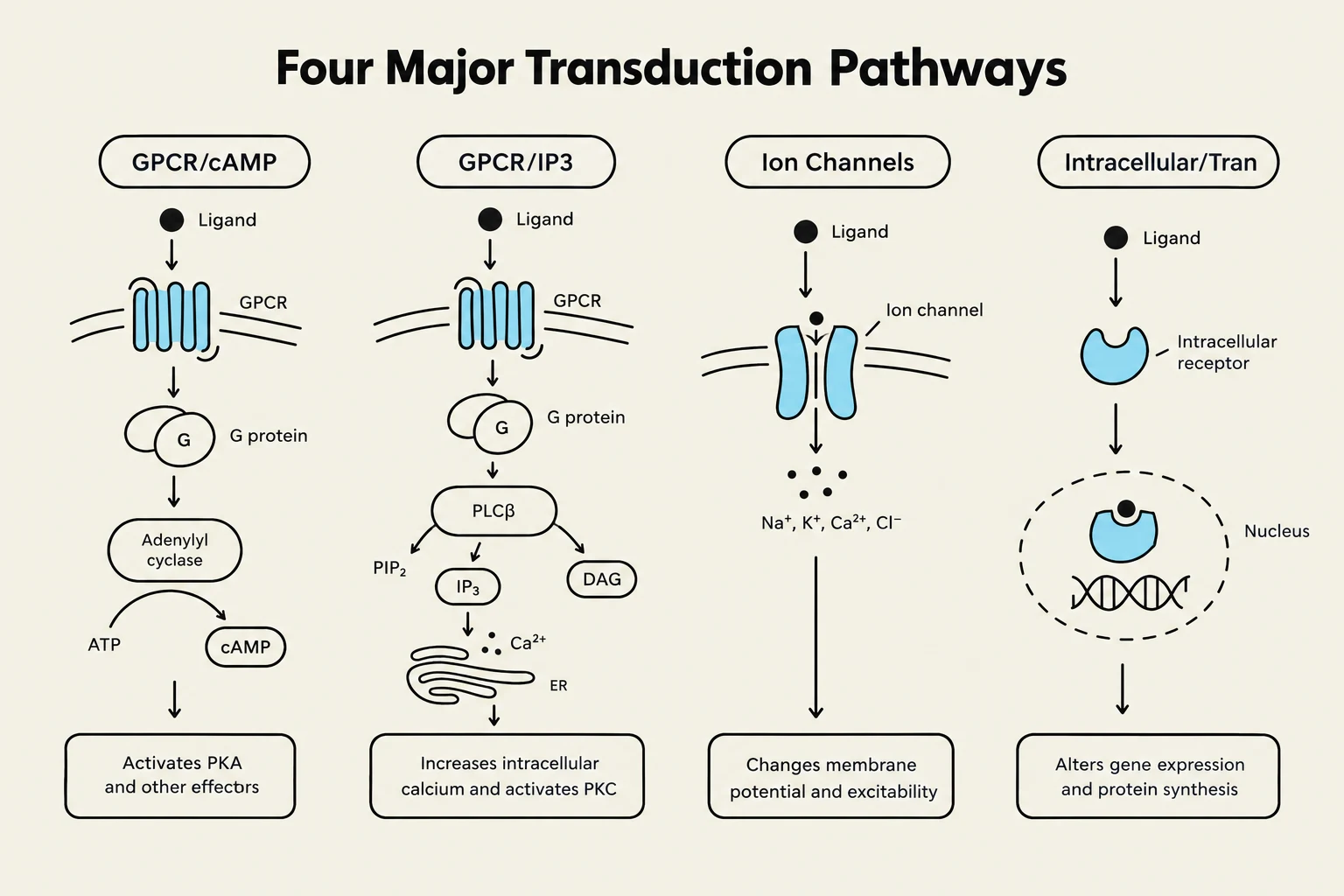
GPCRs represent the largest group of drug-receptor interactions. These receptors possess seven transmembrane domains. When an agonist binds to the extracellular domain, it activates an intracellular G protein, which consists of three subunits: α, β, and γ. The binding of GTP to the α subunit causes the α-GTP complex to dissociate from the βγ complex, allowing them to interact with cellular effectors.
The specific response depends on the type of α subunit:
Gq (αq): Activates phospholipase C, which increases inositol triphosphate (IP3) and diacylglycerol (DAG), leading to increased cytoplasmic Ca2+.
2. Ligand-Gated Ion Channels
These receptors act as gates for ions. When a ligand binds to a specific site on the receptor subunit, it alters the pore size, allowing ions to flow across the membrane. This can result in membrane depolarization. Examples include:
Excitatory: Glutamate or Aspartate receptors.
Inhibitory: GABA or Glycine receptors.
Specific Ion Channels:Na+ channels (inhibited by lidocaine), Ca2+ channels (inhibited by verapamil), and $K^+$ channels (inhibited by amiodarone).
3. Transmembrane Receptor-Effector Enzymes
These receptors (often called catalytic receptors) undergo a conformational change upon ligand binding, typically involving receptor tyrosine kinase activity. This leads to auto-phosphorylation of tyrosine residues on the receptor, which then activates downstream signaling proteins. These responses generally last from minutes to hours.
4. Intracellular Receptors
These receptors are located inside the cell (e.g., in the nucleus). Because the receptor is entirely intracellular, the drug must be lipid-soluble to diffuse across the cell membrane. These drugs often act as transcription factors that regulate gene expression by affecting the transcription of DNA to RNA and subsequently to protein synthesis. Because this process involves protein synthesis, the response takes hours to days to occur. Common examples include steroid hormones.
This diagram illustrates four major signal transduction pathways: GPCR/cAMP, GPCR/IP3, Ion Channels, and Intracellular/Transduction. Each pathway shows the sequence of molecular events triggered by a ligand binding to a receptor, leading to specific cellular responses.
Dose-Response Relationships
The relationship between the concentration of a drug at the receptor site and the magnitude of the resulting response is the dose-response relationship.
Graded vs. Quantal Dose-Response
There are two primary ways to quantify these relationships:
Graded Dose-Response Relationship: This describes the response of an individual. As the dose increases, the response increases until it reaches a plateau (the ceiling effect).
Potency (EC50 or ED50): The concentration or dose required to elicit 50% of the maximal response. A drug with a lower EC50 is more potent because it requires less drug to achieve the same effect.
Efficacy (Emax): The maximal response a drug can produce.
Affinity (Kd): The equilibrium dissociation constant (Kd) is the concentration of drug that binds to 50% of the receptors. Affinity is inversely proportional to Kd (Affinity=Kd1).
Quantal Dose-Response Relationship: This describes the response of a population. It is an "all-or-none" measurement (e.g., did the patient sleep or not?).
ED50 (Median Effective Dose): The dose that produces the desired effect in 50% of the population.
TD50 (Median Toxic Dose): The dose that produces a toxic effect in 50% of the population.
LD50 (Median Lethal Dose): The dose that is lethal to 50% of the population.
Safety Metrics: Therapeutic Index and Window
To assess how safe a drug is, we use the Therapeutic Index (TI), which is the ratio of the toxic dose to the effective dose:
TI=ED50TD50
A large TI indicates a wide margin of safety (e.g., penicillin), whereas a small TI indicates a narrow therapeutic window (e.g., warfarin), requiring careful patient monitoring to avoid toxicity. The therapeutic window is the actual range of doses between the minimum effective concentration and the minimum toxic concentration.
Pharmacodynamics of Agonists and Antagonists
Drugs are further categorized by how they influence receptor activity.
Agonists
An agonist binds to a receptor and activates it, possessing both affinity and efficacy.
Full Agonist: Produces the maximal possible response (Emax).
Partial Agonist: Binds to the receptor but can only produce a submaximal response, even at high doses. For example, buprenorphine is a partial opioid agonist; it cannot achieve the same pain relief as the full agonist morphine.
Inverse Agonist: Stabilizes the receptor in its inactive state, exerting the opposite effect of an agonist.
Allosteric Activator: Binds to a site different from the primary binding site to increase the likelihood that an agonist will bind, effectively increasing potency or efficacy. Benzodiazepines are allosteric activators of the GABA receptor.
Antagonists
An antagonist has affinity (it binds) but zero efficacy (it does not activate the receptor).
Competitive Antagonist: Binds reversibly to the same active site as the agonist. Because it is reversible, you can overcome the inhibition by increasing the concentration of the agonist. This shifts the dose-response curve to the right (increasing EC50/decreasing potency) but does not change the Emax (efficacy). Example: Pravastatin competes with cholesterol for the HMG-CoA reductase enzyme.
Noncompetitive Antagonist: Binds either to the active site irreversibly or to an allosteric site. Increasing the agonist dose cannot overcome this inhibition. This reduces the number of available receptors, shifting the dose-response curve downward (decreasing Emax/efficacy). Example: Aspirin irreversibly inhibits the cyclooxygenase enzyme.
Chemical Antagonist: Interacts directly with the drug itself to neutralize it (e.g., protamine neutralizing heparin).
Functional (Physiological) Antagonist: Acts through a different molecular mechanism to produce an opposite physiological effect (e.g., glucagon counteracting the effects of insulin).
Spare Receptors
In many systems, it is not necessary to occupy every single receptor to achieve a maximal response. This phenomenon is known as having spare receptors. For instance, insulin receptors have a massive functional reserve, where only 1% occupancy is needed for a maximal response. Conversely, the heart has very few spare β-adrenergic receptors, meaning nearly all must be occupied to achieve maximum contractility.
Keep reading for free.
Create an account to finish this reading and make it part of your study routine.