


Perfusion is the life-sustaining process of delivering oxygen, fluids, and nutrients to the cells of the body. Without adequate perfusion, tissues cannot maintain metabolic functions, leading to cellular dysfunction or death. To understand how the body maintains this delicate balance, we must examine the relationship between the heart's mechanical action and the vascular pathways that transport blood throughout the system.
The Mechanics of Perfusion: Pulse and Blood Pressure
Effective perfusion is clinically measured through two primary indicators: pulse and blood pressure.
Pulse represents the blood being pumped from the heart into the systemic circulation in pulsatile waves. It is a tactile reflection of the heart's rhythm and the strength of its contractions.
Blood Pressure (BP) is the force exerted on the arterial walls by the circulating blood. It is not a static number but a dynamic value influenced by the interplay between how much blood the heart pumps and how much resistance the vessels provide.
Mathematically, blood pressure is governed by the relationship between Cardiac Output (CO) and Systemic Vascular Resistance (SVR):
Cardiac Output and Stroke Volume
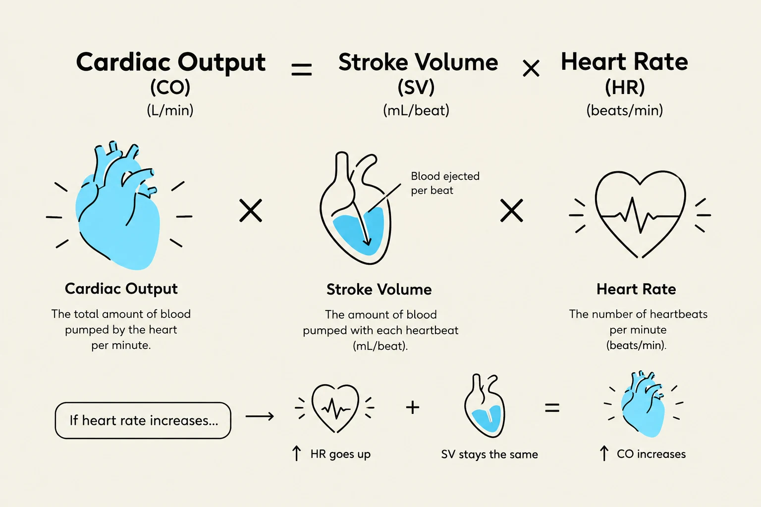
Cardiac Output is the total volume of blood ejected by the heart in one minute. It is calculated using the following formula:
A normal CO typically ranges from 4 to 8 liters per minute. Stroke Volume—the amount of blood pumped during a single contraction—is influenced by three critical factors:
- Preload: The volume of blood in the ventricles at the end of diastole (rest).
- Afterload: The resistance the left ventricle must overcome to circulate blood.
- Contractility: The inherent strength of the myocardial contraction.
Keep reading for free.
Create an account to finish this reading and make it part of your study routine.
Continue reading freeAlready have an account? Log in
Updated