Explains the core concepts of oxygenation and respiratory care, including ventilation, gas exchange, respiratory anatomy, clinical assessment of respiratory distress, and oxygen delivery methods.
Effective respiratory care is a cornerstone of clinical practice, requiring a precise understanding of how the body moves air, exchanges gases, and delivers oxygen to the tissues. To provide safe and effective care, healthcare providers must distinguish between three critical, yet distinct, processes: ventilation (the mechanical movement of air in and out of the lungs), oxygenation (the process of getting oxygen into the blood), and perfusion (the flow of blood through the tissues).
Respiratory Anatomy and Structures
The respiratory system is divided into the upper and lower respiratory tracts. The upper tract includes the nose, nasal cavity, pharynx, and larynx. Air then travels through the trachea into the lower respiratory tract, which consists of the bronchi, bronchioles, and finally, the alveoli—the tiny air sacs where the "magic" of gas exchange happens.
Supporting this entire process are the breathing muscles, most notably the diaphragm and the intercostal muscles, as well as the pleura which protects the lungs.
The Mechanics of Breathing: Ventilation and Gas Exchange
The process of breathing can be broken down into several sequential stages, moving from the macro-level movement of air to the micro-level movement of molecules.
Pulmonary Ventilation
Ventilation is the physical movement of air. This involves two phases:
Inspiration (Inhalation): An active process where the diaphragm contracts to draw air in through the nose or mouth and down the airways to the alveoli.
Expiration (Exhalation): A passive process where the diaphragm relaxes, allowing carbon dioxide to leave the body.
Alveolar Gas Exchange
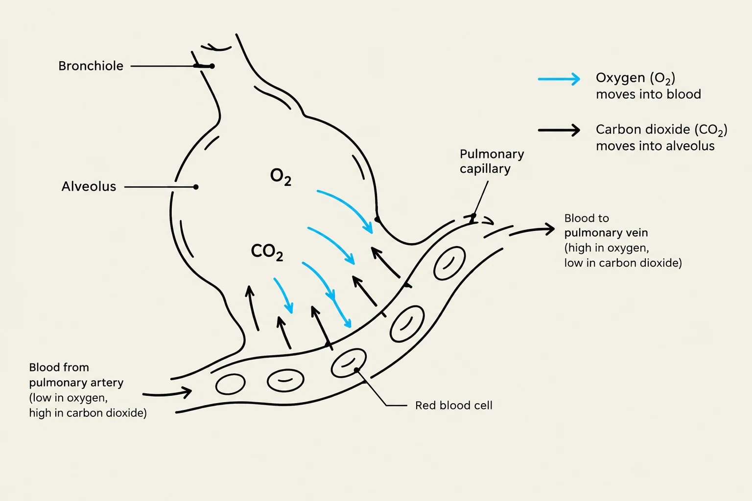
Once air reaches the alveoli, gas exchange occurs via diffusion. Oxygen moves from the high concentration in the alveoli into the pulmonary blood, while carbon dioxide moves from the blood into the alveoli to be exhaled.
Diagram illustrating gas exchange in the alveoli of the lungs, showing oxygen moving from the alveoli into the pulmonary capillaries and carbon dioxide moving from the capillaries into the alveoli.
Gas Transport and Tissue Exchange
After oxygen enters the blood, it must be transported to the rest of the body. Oxygen binds to the hemoglobin within red blood cells (RBCs), which act like "delivery trucks". Once the blood reaches the systemic capillaries near the body's cells, tissue gas exchange occurs: oxygen leaves the blood to enter the cells, and carbon dioxide moves from the cells into the blood to be carried away.
When assessing a patient, clinicians must look beyond just the numbers. A comprehensive respiratory assessment includes observing the rate, depth, rhythm, and effort of breathing, as well as chest movement.
Characteristics of Breathing
Rate: Measured in breaths per minute. Abnormalities include tachypnea (fast), bradypnea (slow), or apnea (absence of breathing).
Depth: Can range from shallow to deep.
Effort: This refers to the work required to breathe. Signs of distress include dyspnea (shortness of breath), nasal flaring, retractions, or the use of accessory muscles.
Abnormal Findings
Clinicians should listen for adventitious breath sounds, which are abnormal sounds such as wheezes, crackles, stridor, or rhonchi. Another critical sign is cyanosis, a bluish discoloration of the skin indicating low oxygen levels.
Exam Tip: Always remember to compare lung sounds bilaterally (on both sides) to ensure symmetry in ventilation.
Understanding Oxygenation: Hypoxemia vs. Hypoxia
It is a common clinical error to use "hypoxia" and "hypoxemia" interchangeably, but they refer to different physiological states.
Hypoxemia: Refers specifically to low oxygen levels in the arterial blood (low PaO2).
Hypoxia: Refers to inadequate oxygen reaching or being used by the tissues.
Hypoxia can be caused by hypoxemia, but it can also be caused by poor circulation (shock), anemia (not enough hemoglobin to carry oxygen), or impaired cellular use of oxygen.
Ventilation-Perfusion (V/Q) Mismatch
Respiratory efficiency relies on the balance between ventilation (V) and perfusion (Q).
Shunt: Occurs when blood flows past alveoli that are not being ventilated (e.g., due to pneumonia or pulmonary edema).
Dead Space: Occurs when air reaches the alveoli, but there is little to no blood flow in the nearby capillaries (e.g., a pulmonary embolism).
Monitoring and Oxygen Delivery
Pulse Oximetry (SpO2)
Pulse oximetry estimates the percentage of hemoglobin saturated with oxygen. However, it has significant limitations:
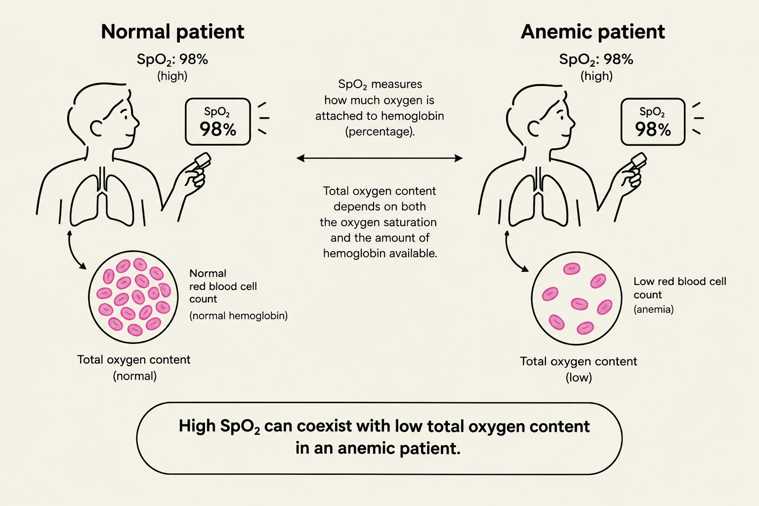
It does not measure total hemoglobin levels. An anemic patient may have a normal SpO2 (percentage of seats filled) even if they have very few "seats" (hemoglobin) available.
It does not measure CO2 levels or actual tissue oxygenation.
Accuracy can be affected by poor perfusion, cold fingers, nail polish, or excessive movement.
This illustration compares oxygen saturation (SpO2) and total oxygen content in a normal patient versus an anemic patient. Both patients can have a high SpO2 of 98%, but the anemic patient has a low total oxygen content due to a reduced red blood cell count, which carries hemoglobin. This highlights that high SpO2 does not always equate to high total oxygen content.
Oxygen Delivery Devices
Oxygen is delivered via various devices depending on the patient's needs:
Device
Flow Rate
Purpose/Notes
Nasal Cannula
2–6 L/min
Low flow via nasal prongs
Simple Face Mask
6–10 L/min
Covers nose and mouth
Venturi Mask
Variable
Delivers precise, controlled concentrations
Nonrebreather Mask
10–15 L/min
High flow; keep reservoir inflated
For more advanced support, clinicians use High-Flow Nasal Cannula (HFNC), CPAP (continuous positive airway pressure), BiPAP (two levels of pressure), or mechanical ventilation.
Medication Delivery and Nursing Priorities
Inhalation Therapy
Medications are often delivered directly to the lungs to improve effectiveness:
Nebulizers: Turn liquid medication into a mist. Patients should sit upright and breathe normally until the mist stops.
Metered Dose Inhalers (MDI): Handheld aerosol devices. Using a spacer with an MDI is highly recommended as it helps more medication reach the lungs rather than just hitting the back of the throat.
Nursing Safety and Responsibilities
When managing oxygen therapy, nursing priorities include:
Verification: Always verify the provider's order for the specific device and flow rate.
Assessment: Assess the patient both before and after changing oxygen settings.
Safety: Oxygen is a fire accelerant. Keep it away from flames, heat, and smoking.
Skin Integrity: Regularly check the skin where tubing or masks contact the patient to prevent breakdown.
Keep reading for free.
Create an account to finish this reading and make it part of your study routine.