An explanation of the physical principles behind Magnetic Resonance Imaging (MRI), covering atomic properties, magnetic fields, resonance, relaxation, and signal detection for image contrast.
Magnetic Resonance Imaging (MRI) is a sophisticated medical imaging technique that allows clinicians to see detailed, cross-sectional images of the human body without using ionizing radiation. Unlike X-rays or CT scans, MRI relies on the magnetic properties of atoms—specifically hydrogen—to create contrast between different biological tissues.
To understand how an MRI scanner transforms a patient's body into a digital image, we must follow a sequence of physical events: alignment with a magnetic field, energy absorption through resonance, and the subsequent detection of signals as those atoms return to their natural state.
The Building Block: Hydrogen Protons
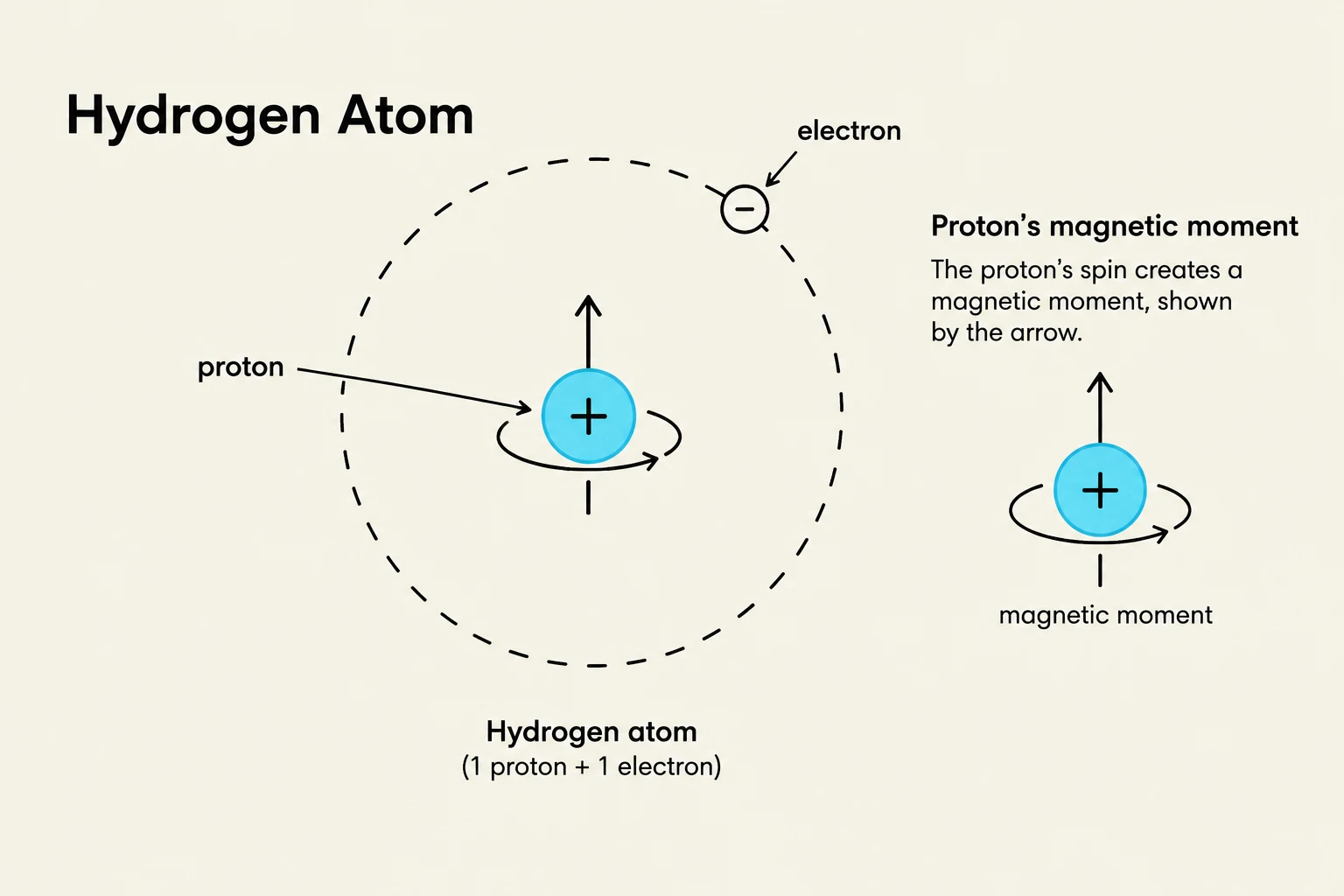
The foundation of MRI is the hydrogen atom. A hydrogen atom consists of a single proton in its nucleus and one electron orbiting outside. For the purposes of MRI, we are primarily interested in the proton because it possesses a positive charge and a magnetic property that allows it to act like a tiny magnet.
Hydrogen is the ideal target for imaging because it is incredibly abundant in the human body, found in high concentrations within water and fat. This abundance ensures that there are plenty of "tiny magnets" available to provide a strong, detectable signal.
Diagram illustrating the structure of a hydrogen atom, showing a central proton with a positive charge and an orbiting electron with a negative charge. It also depicts the proton's magnetic moment.
Alignment and the Net Magnetization Vector
In their natural state, outside of a scanner, these hydrogen protons spin in random directions. Because they point every which way, their individual magnetic effects cancel each other out.
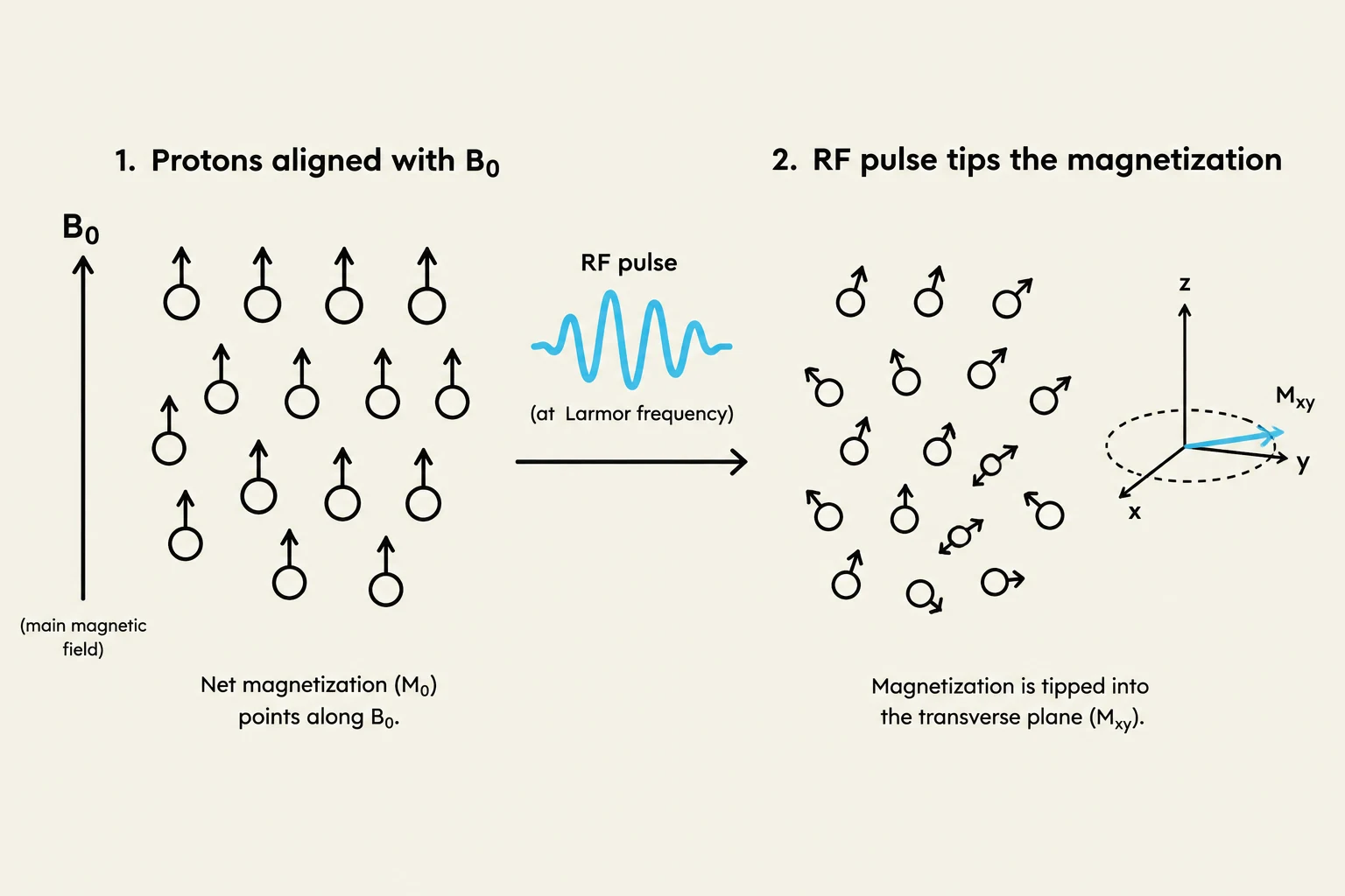
However, when a patient is placed inside the scanner, they are exposed to a powerful external magnetic field known as B0 (the main magnetic field). Under the influence of B0, the protons begin to align themselves. They can align either parallel (pointing in the same direction as the field) or anti-parallel (pointing in the opposite direction).
Because it requires less energy to align parallel to the field, a slight majority of protons will adopt this orientation. This slight imbalance creates an overall magnetic effect called the net magnetization vector.
Exam Tip: Remember that the net magnetization vector is not a result of all protons pointing one way, but rather the difference between the number of protons pointing parallel versus anti-parallel to B0.
Resonance and Excitation
Once the protons are aligned, the system must "disturb" them to elicit a signal. This is achieved by applying radiofrequency (RF) energy.
To make the protons absorb this energy, the RF pulse must be applied at a very specific frequency that matches the protons' own frequency. This phenomenon is called resonance. A common way to visualize this is the "swing analogy": if you push a person on a swing, you must time your pushes to match the rhythm of the swing to transfer energy effectively; if your timing is off, you won't move them much.
When resonance occurs, there is a successful energy transfer. This causes the net magnetization vector to "flip" or change direction away from the B0 axis.
This illustration depicts the process of nuclear magnetic resonance (NMR) where protons align with a magnetic field (B0) and are then tipped into the transverse plane by an RF pulse at the Larmor frequency, resulting in magnetization in the transverse plane (Mxy).
Relaxation: T1 and T2
After the RF pulse is turned off, the protons are no longer being "pushed." They want to return to their original, low-energy state. This process is known as relaxation. Relaxation occurs through two simultaneous but independent processes:
T1 Recovery: This is the process where the magnetization builds back up along the direction of the main magnetic field (B0).
T2 Decay: This is the process where the protons lose their coordination (or "phase") with one another, causing the signal to decrease over time.
While these two processes happen at the same time, they describe different physical changes in the tissue.
Signal Detection and Image Contrast
As the protons relax, they release the energy they previously absorbed. We need a way to "catch" this energy to turn it into an image. This is the job of the receiver coils. You can think of these coils as antennas placed near the specific body part being scanned (such as a head coil or a knee coil).
The physics behind this detection is explained by Faraday’s Law, which states that a changing magnetic field induces an electrical signal in a conductor (the coil). As the relaxing protons create a changing magnetic environment, the coil picks up the resulting electrical signal.
The final step is creating the image. Different biological tissues (like muscle, fat, or water) have different rates of T1 recovery and T2 decay. Because they relax at different speeds, they produce different signal strengths. This difference in signal strength is what provides image contrast, allowing a radiologist to distinguish between a tumor, a cyst, or healthy tissue on the screen.
Process
Physical Action
Resulting Effect
T1 Recovery
Magnetization regrows along B0
Longitudinal signal recovery
T2 Decay
Protons lose coordination
Transverse signal loss
Summary Checklist for MRI Physics:
Magnetic: B0 aligns the protons.
Resonance: RF energy excites the protons at a specific frequency.
Imaging: Receiver coils detect the signal during relaxation to create an image.
Keep reading for free.
Create an account to finish this reading and make it part of your study routine.