Fundamentals of Immunogenicity and Antigen Presentation
Explores the core principles of how the immune system distinguishes self from non-self, defines antigens, and presents them to immune cells using MHC proteins and T-cell receptors.
To understand how the immune system protects the body, one must first understand how it distinguishes "self" from "non-self". The immune system does not simply react to everything it encounters; rather, it relies on a sophisticated recognition system that identifies specific molecular patterns. This article explores the fundamental principles of how antigens are defined, how they are presented to immune cells, and the molecular machinery—specifically MHC proteins and T-cell receptors—that makes this recognition possible.
Immunogenicity and Antigenicity
While the terms are often used interchangeably in casual conversation, they have distinct meanings in immunology. Antigenicity refers to the ability of a substance to bind specifically to the products of an immune response, such as antibodies or T-cell receptors. Immunogenicity, however, is a more rigorous property: it is the ability of a substance to induce an immune response in the first place.
Not all antigens are immunogens. For example, a hapten is a small molecule that possesses antigenicity (it can bind to an antibody) but lacks immunogenicity (it cannot trigger an immune response on its own). Haptens can only become immunogenic if they attach to a larger "carrier" protein.
The Nature of Epitopes
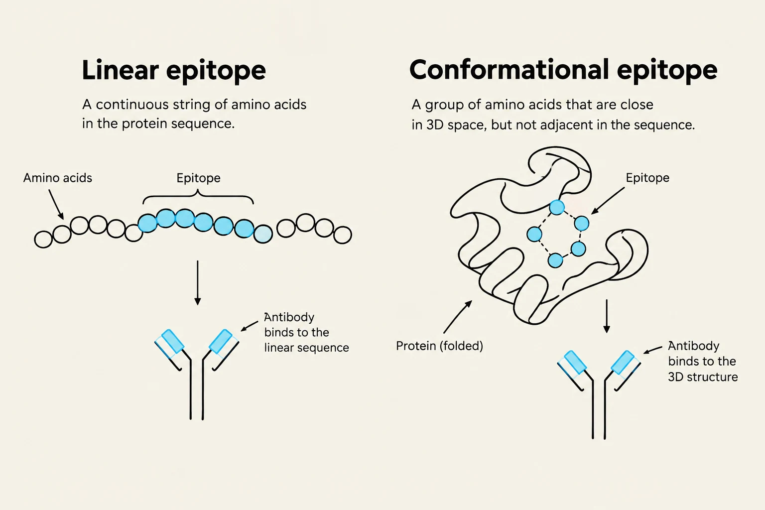
The specific part of an antigen that is recognized by the immune system is called an epitope or antigen determinant. Epitopes generally fall into two categories:
Linear epitopes: Created by a continuous sequence of amino acids in a polypeptide chain.
Conformational epitopes: Created by the folding of a protein, bringing distant amino acid residues into close proximity.
Exam Tip: Remember that B-cells can often recognize conformational epitopes on the surface of intact proteins, whereas T-cells typically require processed, linear peptides presented on MHC molecules.
This illustration differentiates between linear and conformational epitopes, explaining how antibodies bind to continuous amino acid sequences or to specific 3D structures on proteins, respectively.
Classifying Antigens
Antigens can be classified based on their relationship to the host:
Autoantigens: Molecules that are part of the "self" and should ideally be ignored by the immune system.
Alloantigens: Molecules that are "self" to one individual but "foreign" to another of the same species (e.g., MHC molecules in organ transplants).
Heteroantigens: Molecules that are entirely foreign to the species (e.g., bacterial or viral proteins).
When the immune system mistakenly attacks autoantigens, it leads to autoimmunity. This can sometimes be triggered by cross-reactivity, where an immune response against a foreign antigen (like a virus) inadvertently attacks a self-antigen because they share similar epitopes.
To enhance the immune response during vaccination, scientists often use adjuvants—substances added to an antigen to increase its immunogenicity.
MHC Proteins: The Display Window
T-cells are unique because they cannot "see" free-floating antigens in the blood or tissues. Instead, they can only recognize antigens that have been processed and "presented" on the surface of other cells via Major Histocompatibility Complex (MHC) proteins.
The MHC serves as a molecular display window. By presenting internal or external proteins on the cell surface, the MHC allows the immune system to survey the "health" of the cell. The genetics of the MHC are highly complex and polymorphic, meaning there is immense variation in MHC molecules between individuals. This variation is the biological reason why some people are more susceptible to certain diseases or why organ rejection occurs.
MHC Class I and II Structure
There are two primary classes of MHC molecules involved in antigen presentation:
MHC Class I: Found on almost all nucleated cells. They present endogenous antigens (proteins produced inside the cell, such as viral proteins) to CD8+ cytotoxic T-cells.
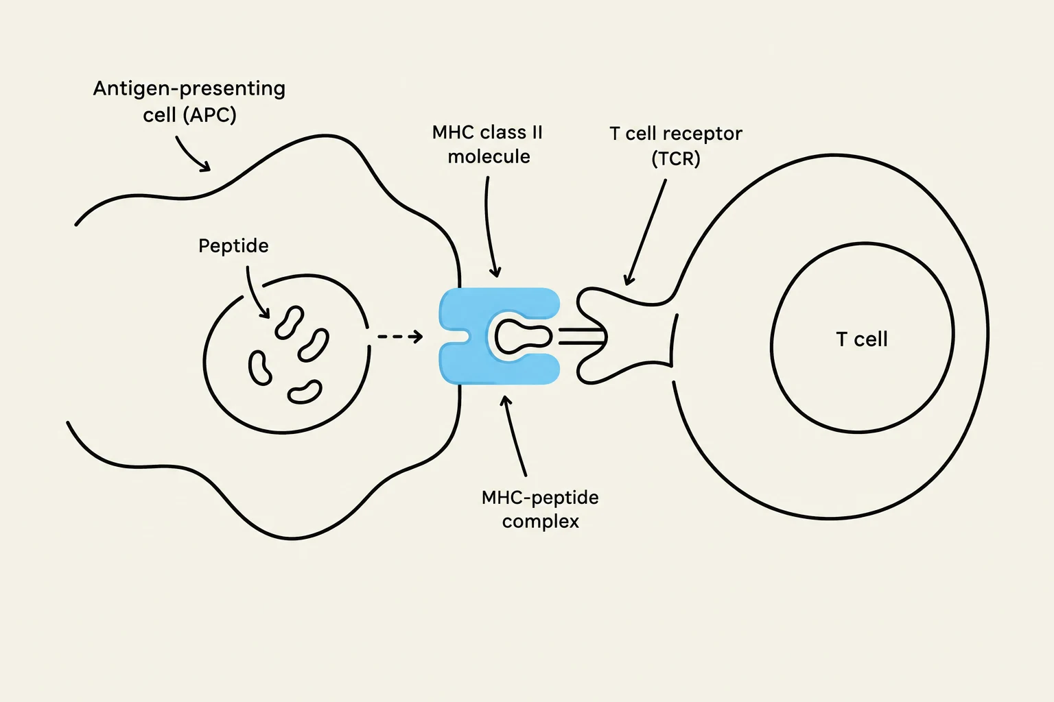
MHC Class II: Found primarily on specialized Antigen-Presenting Cells (APCs) like macrophages, B-cells, and dendritic cells. They present exogenous antigens (proteins taken up from outside the cell) to CD4+ helper T-cells.
Diagrams illustrating the structure of MHC Class I and MHC Class II molecules, showing their peptide-binding grooves and association with cell membranes.
T-Cell Receptors (TCR)
The T-cell receptor (TCR) is the molecule responsible for "reading" the MHC-antigen complex. The TCR is a membrane-bound protein consisting of two chains (usually alpha and beta).
Gene Rearrangement and Diversity
To ensure that the body can recognize virtually any possible pathogen, T-cells undergo a process called gene rearrangement. During T-cell development, the genes encoding the TCR chains are shuffled and recombined. This creates a massive repertoire of unique receptors, ensuring that even though each T-cell has only one specific receptor, the total population of T-cells can recognize almost any epitope.
Signaling and the Zeta Subunits
The TCR itself has a very short cytoplasmic tail, meaning it cannot send signals into the cell on its own. To solve this, the TCR is non-covalently associated with signaling subunits, most importantly the zeta (ζ) subunits. When the TCR binds to an MHC-antigen complex, these zeta subunits transmit the biochemical signal into the T-cell, triggering activation and proliferation.
Antigen Presentation and APC-T Cell Interactions
The final step in the recognition process is the physical interaction between an Antigen-Presenting Cell (APC) and a T-cell.
In a typical interaction:
An APC (like a dendritic cell) engulfs a pathogen.
The pathogen is broken down into peptides.
These peptides are loaded onto MHC molecules.
The APC moves to a lymph node where it encounters a T-cell with a matching TCR.
The stability of this interaction is crucial. The T-cell must recognize both the peptide (the antigen) and the specific "shape" of the MHC molecule itself. This "dual recognition" ensures that T-cells only respond to antigens presented in the correct biological context.
Diagram illustrating the interaction between an antigen-presenting cell (APC) and a T cell. The APC presents a peptide fragment via an MHC class II molecule, which is recognized by the T cell receptor (TCR) on the T cell.
Summary Table: Antigen Presentation Pathways
Feature
MHC Class I
MHC Class II
Cell Type
All nucleated cells
Professional APCs
Antigen Source
Endogenous (Intracellular)
Exogenous (Extracellular)
Target T-cell
CD8+ Cytotoxic T-cells
CD4+ Helper T-cells
Function
Identifies infected/cancerous cells
Coordinates immune response
Keep reading for free.
Create an account to finish this reading and make it part of your study routine.