Explores the fundamental principles of DNA structure, replication, transcription, and the organization of genetic material within cells, including mitosis and meiosis.
Understanding the molecular basis of life requires a deep dive into the structures that store, replicate, and transmit genetic information. From the chemical polarity of a single nucleic acid strand to the complex dance of chromosomes during meiosis, the precision of these processes ensures biological continuity. This article explores the fundamental principles of DNA structure, the mechanics of replication and transcription, and the organizational hierarchy of the cell.
The Chemical Structure of Nucleic Acids
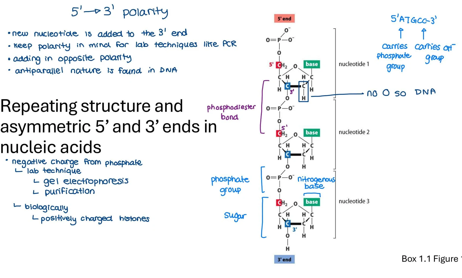
At the most basic level, DNA is composed of nucleotides linked by phosphodiester bonds. A critical feature of these strands is their 5' to 3' polarity. This asymmetry means that new nucleotides are always added to the 3' end of a growing strand. This polarity is a vital consideration in laboratory techniques such as PCR (Polymerase Chain Reaction), where primers must be designed with the correct orientation.
Diagram illustrating the 5' to 3' polarity and repeating structure of nucleic acids, including the components of nucleotides and phosphodiester bonds.Figure 1: DNA polarity and phosphodiester bonds.
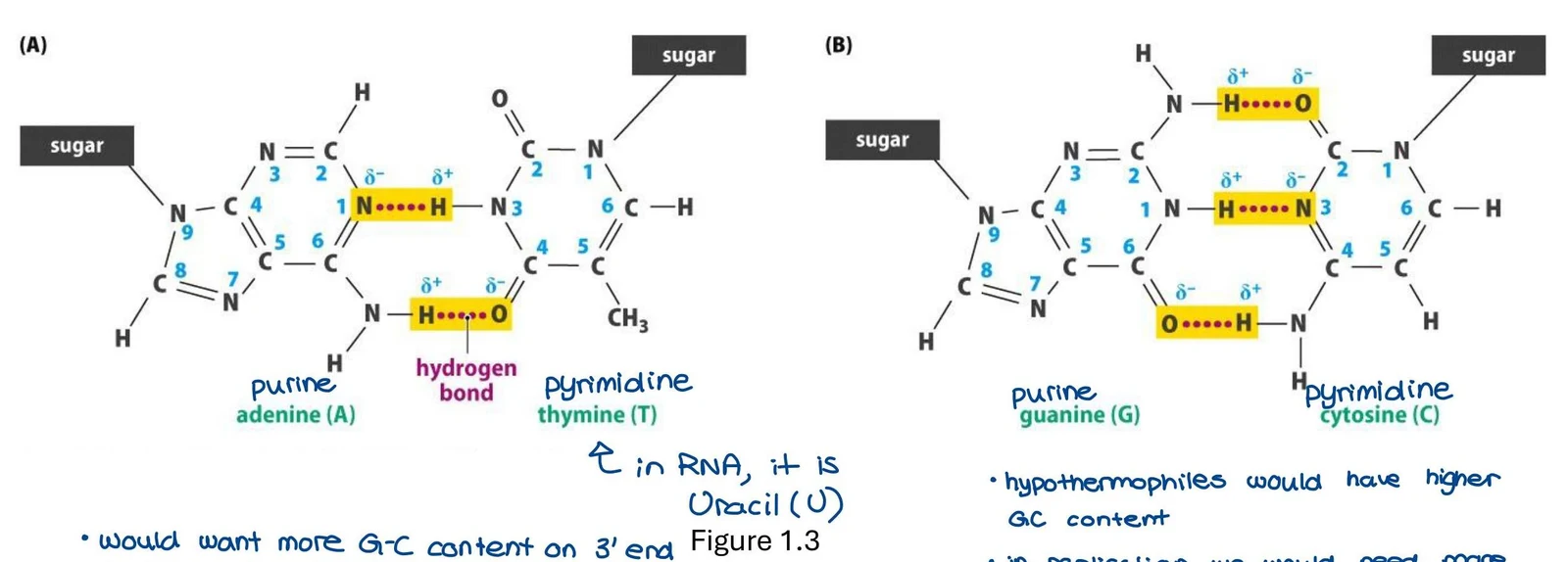
DNA is characterized by its antiparallel nature, meaning the two strands of the double helix run in opposite directions. The stability and specificity of this structure are maintained through complementary base pairing. Adenine (A) pairs with Thymine (T), and Guanine (G) pairs with Cytosine (C) via hydrogen bonding.
Diagrams illustrating the base pairing of adenine with thymine (A) and guanine with cytosine (B) in DNA, showing hydrogen bonds and partial charges. It also notes that uracil replaces thymine in RNA and mentions GC content in relation to extremophiles.Figure 1.3: Hydrogen bonding in base pairs.
Study Tip: The G-C content of a DNA sequence affects its stability. Because G-C pairs have more hydrogen bonds, sequences with high G-C content are more stable. For example, hyperthermophiles (organisms living in extreme heat) often have higher GC content to prevent DNA denaturation.
DNA Replication and Polymerases
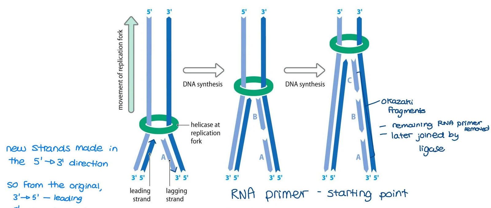
Because of the antiparallel structure and the requirement that DNA polymerases only work in one direction, replication is semi-discontinuous. While one strand (the leading strand) is synthesized continuously, the other (the lagging strand) must be synthesized in short, discontinuous segments known as Okazaki fragments.
Diagram illustrating DNA replication, showing the leading and lagging strands, helicase activity, Okazaki fragments, and the role of RNA primers.Figure 1.5: Semi-discontinuous replication.
Different specialized enzymes, known as DNA polymerases, carry out specific tasks in mammalian cells:
DNA Polymerase
Primary Role
α (alpha)
Initiates DNA synthesis and primes Okazaki fragments
δ (delta) & ε (epsilon)
Major nuclear polymerases; involved in replication and repair
β (beta)
Involved in base excision repair
γ (gamma)
Dedicated to mitochondrial DNA synthesis and repair
TERT (Telomerase)
Replicates the ends of linear chromosomes using an RNA template
Unlike nuclear DNA, which is replicated exactly once per cell cycle, mitochondrial DNA replication is not strictly tied to a single cycle and can occur multiple times.
Transcription and Chromatin Organization
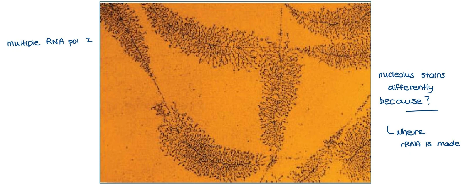
Genetic information is expressed through RNA transcription, where a DNA template is used to synthesize RNA. While mRNA (which codes for proteins) makes up only 1-5% of the total RNA, the majority of transcribed RNA is rRNA (ribosomal RNA). In the nucleolus, multiple RNA Polymerase I molecules work simultaneously to transcribe rRNA, often appearing as "Christmas tree" structures under an electron microscope.
Electron micrograph showing multiple RNA polymerase I molecules transcribing DNA, likely related to rRNA synthesis in the nucleolus.Figure: RNA transcription "Christmas tree" structures.
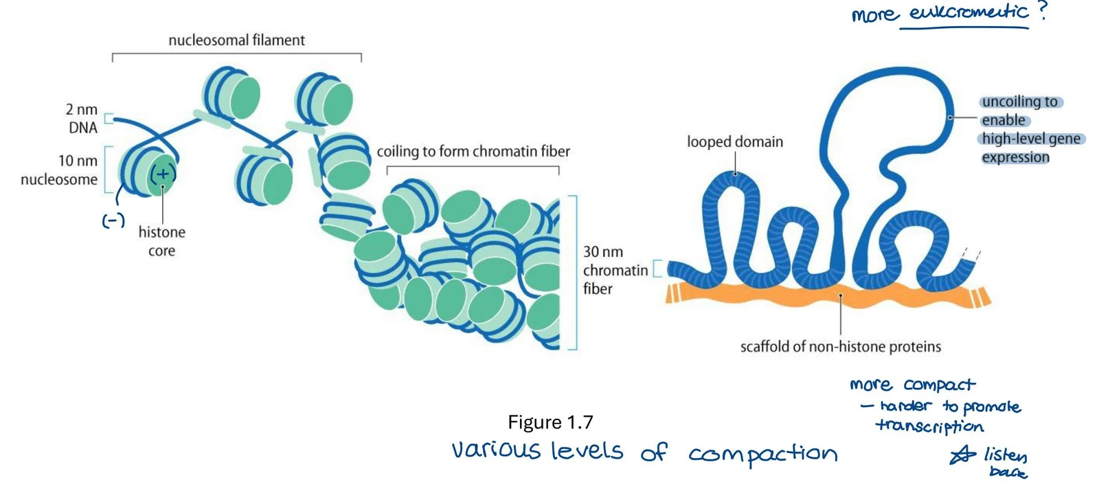
To fit within the nucleus, DNA undergoes hierarchical compaction into interphase chromatin. This organization involves wrapping DNA around histones to form nucleosomes, which then fold into higher-order looped domains.
Diagrams illustrating the levels of DNA compaction in eukaryotic cells, from nucleosomal filaments to looped domains, and their relation to gene expression.Figure: Hierarchical DNA compaction.
Chromosome Dynamics and the Cell Cycle
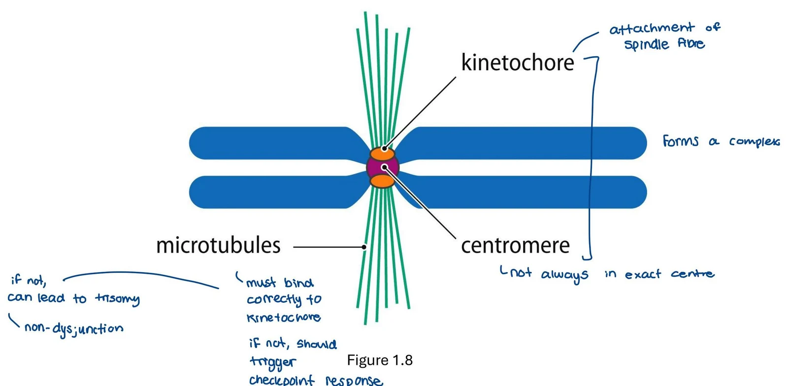
During cell division, chromosomes must be accurately segregated. This relies on the centromere, where kinetochores assemble to attach to microtubules.
Diagram illustrating the attachment of spindle fibers to the kinetochore and centromere of a chromosome, and the consequences of incorrect binding.Figure: Kinetochore and microtubule attachment.
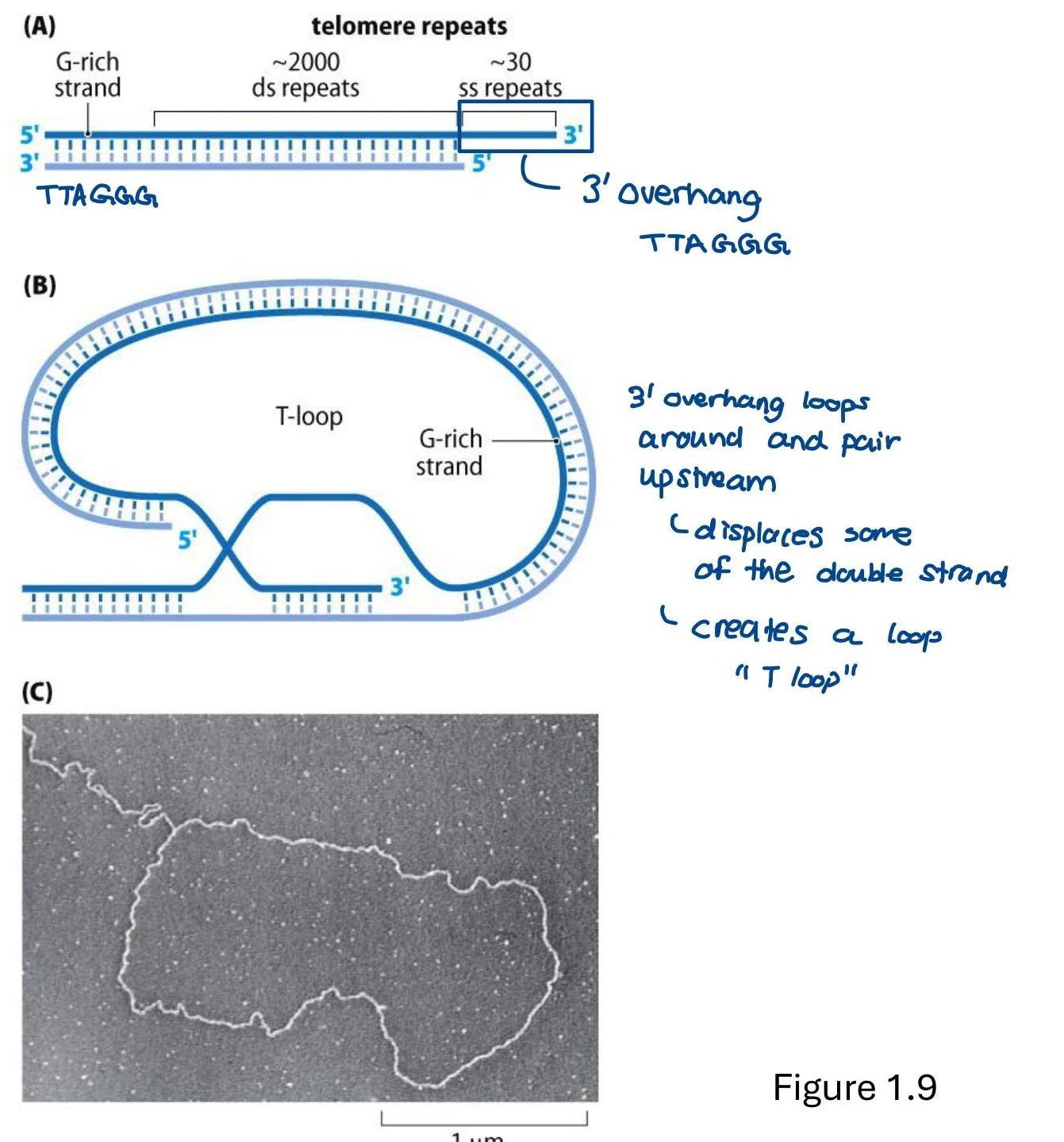
A major challenge for linear chromosomes is protecting the ends from degradation or fusion. This is achieved by telomeres, which form specialized T-loop structures to "hide" the free ends of the DNA. Without these loops, chromosomes could undergo end-to-end fusions, leading to genomic instability.
Diagrams illustrating telomere structure and T-loop formation, with a corresponding electron micrograph.Figure: Telomere T-loop formation.
Mitosis vs. Meiosis
The cell cycle manages the distribution of DNA through two distinct processes:
Mitosis and Cytokinesis: This process results in two genetically identical daughter cells. It is used for growth and tissue repair.
Meiosis: This specialized division produces gametes (sperm or egg cells) for sexual reproduction.
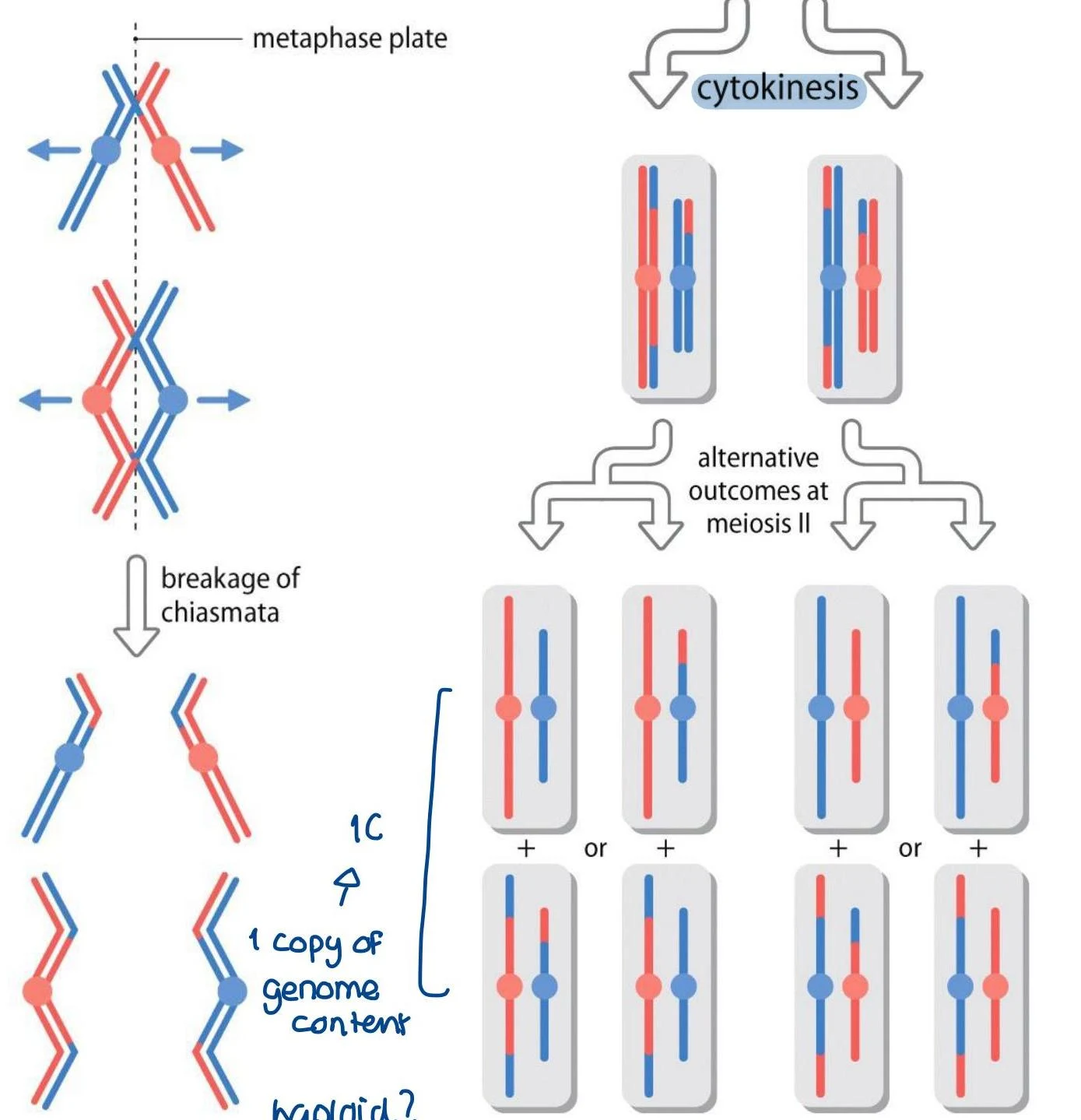
Meiosis introduces massive genetic diversity through two main mechanisms:
Independent Assortment: During Metaphase I, maternal and paternal homologous chromosomes align randomly. This means a single sperm cell might receive a mix of maternal and paternal chromosomes (e.g., a mix of X and Y or different autosomal combinations).
Recombination (Crossing Over): During Prophase I, homologous chromosomes exchange segments of DNA, creating entirely new combinations of alleles that did not exist in either parent.
Diagram illustrating the process of meiosis, including metaphase plate alignment, chiasmata breakage, cytokinesis, and alternative outcomes at meiosis II, showing chromosome segregation and genome content.Figure 1.14: Meiosis and gamete production.
Summary Table: DNA Content and Chromosomes
During the cell cycle, the amount of DNA and the number of chromosomes change dynamically. DNA replication occurs during the S-phase, doubling the DNA content before the cell proceeds through mitosis or meiosis.
Figure: Changes in DNA and chromosome number.
Keep reading for free.
Create an account to finish this reading and make it part of your study routine.