Explore the dynamic roles of membrane proteins in enzymatic activity, structural support, and cell recognition. Understand the critical concept of membrane fluidity and how cells regulate it through fatty acid tails and cholesterol.
The plasma membrane is far more than just a static barrier separating the inside of a cell from the outside world. It is a dynamic, living interface composed of a phospholipid bilayer that serves as a stage for a wide variety of specialized proteins. These membrane proteins are the primary drivers of a cell's specific capabilities; essentially, the types of proteins embedded in a membrane reflect the specific functions that the cell is designed to perform.
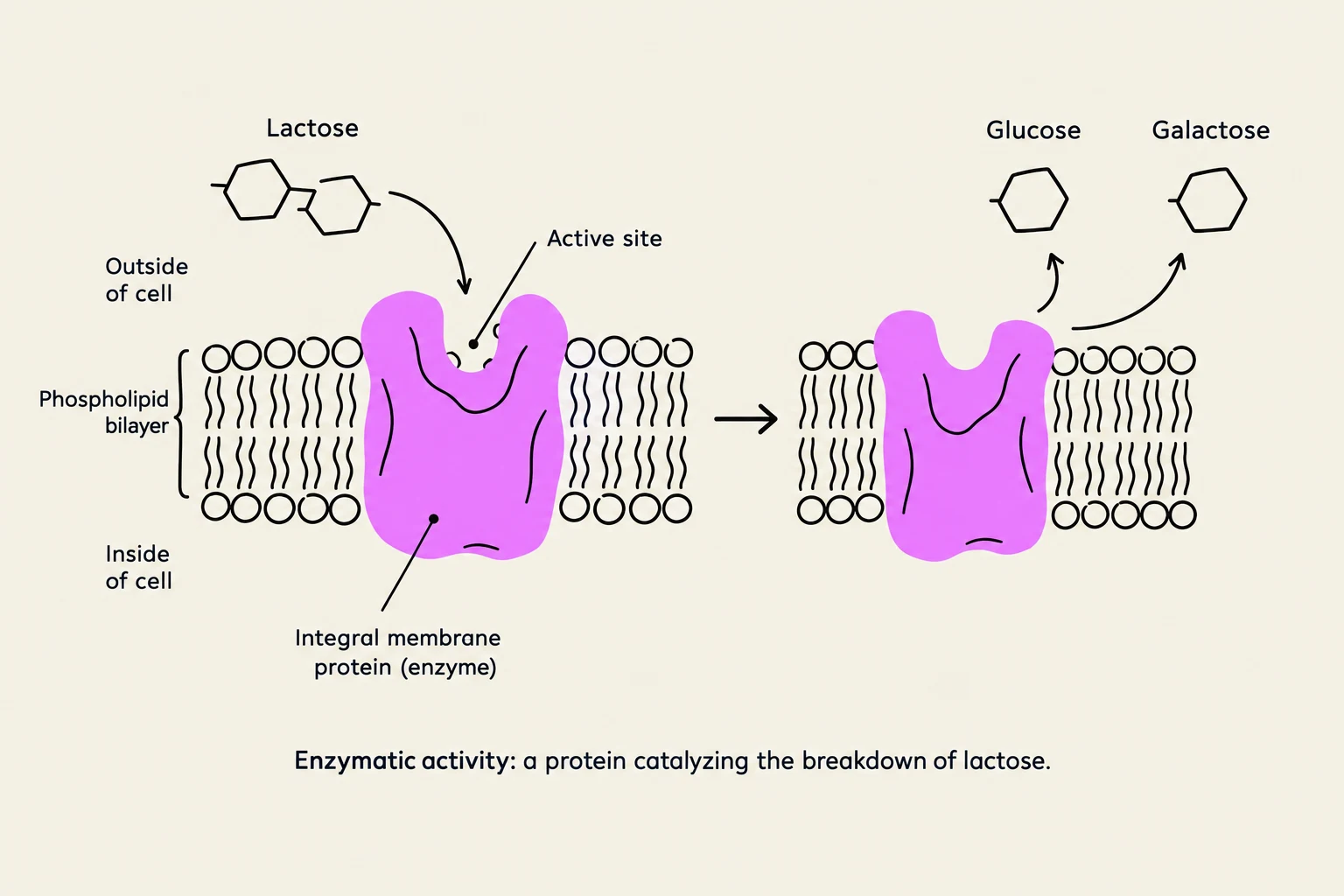
Diagram illustrating the enzymatic breakdown of lactose into glucose and galactose by an integral membrane protein (enzyme) within a phospholipid bilayer.
The Diverse Roles of Membrane Proteins
Because proteins can take on many different shapes and chemical properties, they can perform several distinct roles within the membrane. These functions can be categorized into three primary areas: enzymatic activity, structural support, and cell recognition.
FIGURE 3.3: Functions of membrane proteins.
1. Enzymatic Activity and Catalysis
Some membrane proteins act as enzymes, which are biological catalysts that speed up chemical reactions. These proteins often feature active sites—recessed pockets or clefts on the protein's surface where specific molecules can bind to undergo a transformation.
A classic biological example is the enzyme lactase. Found in the epithelial cells lining the small intestine, lactase facilitates the breakdown of the disaccharide lactose (the sugar found in milk) into simpler, absorbable sugars.
2. Linkers and Structural Support
Cells require internal and external stability to maintain their shape and to interact with their environment. Both integral proteins (which span the membrane) and peripheral proteins (which are attached to the surface) can act as linkers.
By anchoring filaments both inside and outside the plasma membrane, these proteins provide essential structural support. This anchoring can help maintain cell shape, facilitate cell movement, or even link adjacent cells together to form tissues.
3. Cell Recognition and Identity
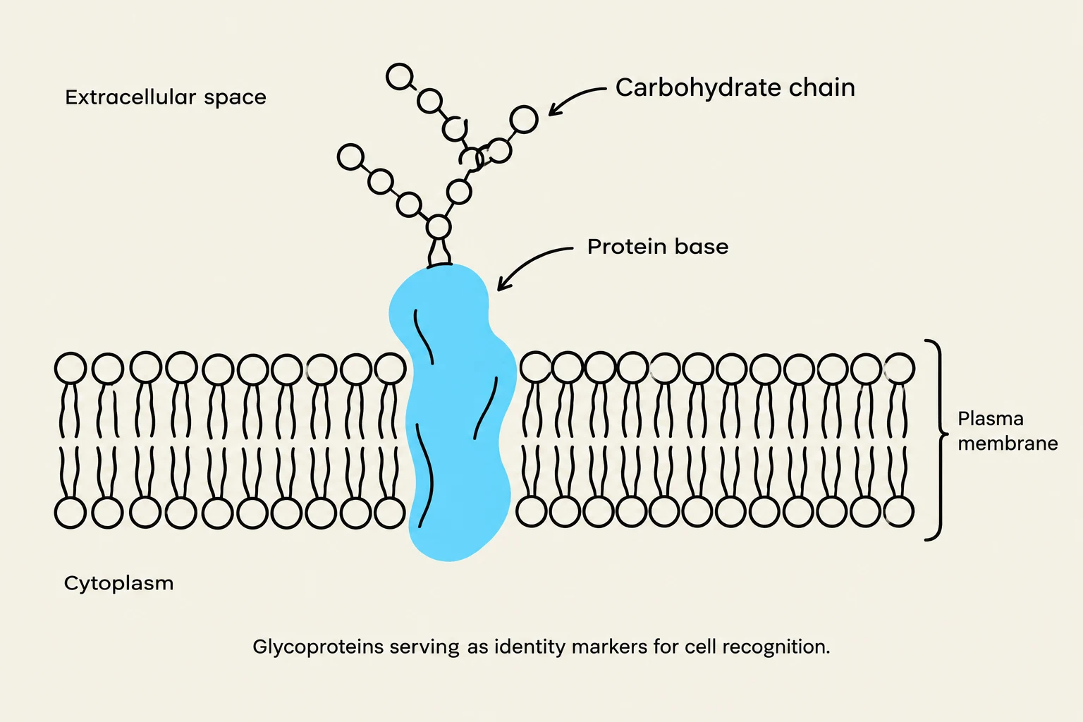
Glycoproteins, composed of a protein base and a carbohydrate chain, serve as identity markers for cell recognition on the plasma membrane.
How does your immune system know which cells belong to you and which are invading bacteria? The answer lies in cell recognition. Many membrane proteins are decorated with carbohydrate chains, forming structures known as glycoproteins.
These glycoproteins act as "identity markers," allowing the body to distinguish its own cells from foreign ones. A critical class of these markers is the major histocompatibility (MHC) proteins, which are vital for immune system function.
Study Tip: Test Your Knowledge
When a hormone like insulin binds to a protein in the plasma membrane to trigger a cellular response, which function is being demonstrated?
Answer: This represents a signaling/receptor function (often categorized under the broader umbrella of protein-mediated interactions), though it is distinct from the three primary categories shown in Figure 3.3.
Membrane Fluidity: The "Fluid Mosaic" in Motion
The plasma membrane is not a solid, frozen sheet; it is a fluid structure. Most membrane lipids and many proteins can rotate and move laterally (sideways) within the bilayer. To give you a sense of the scale, neighboring lipid molecules can exchange places approximately 10 million times per second.
Why Fluidity Matters
Fluidity is a delicate balance that the cell must maintain. If the membrane were too rigid, the proteins wouldn't be able to move to interact with one another; if it were too fluid, the cell would lose its structural integrity.
Proper fluidity enables several critical processes:
Protein Assembly: Allowing proteins to move and cluster together to form functional complexes.
Cellular Processes: Facilitating cell growth, division, movement, secretion, and the formation of cellular junctions.
Self-Sealing: One of the most remarkable properties of the lipid bilayer is its ability to self-seal if it is punctured.
Real-World Application: Medical and Scientific Breakthroughs
Because membranes self-seal, scientists can use thin needles to puncture a cell and then withdraw the needle without the cell bursting. This property makes intracytoplasmic sperm injection (used to help infertile couples) possible, and it allowed for the nuclear transfer experiments used to create Dolly the sheep.
Regulating Fluidity
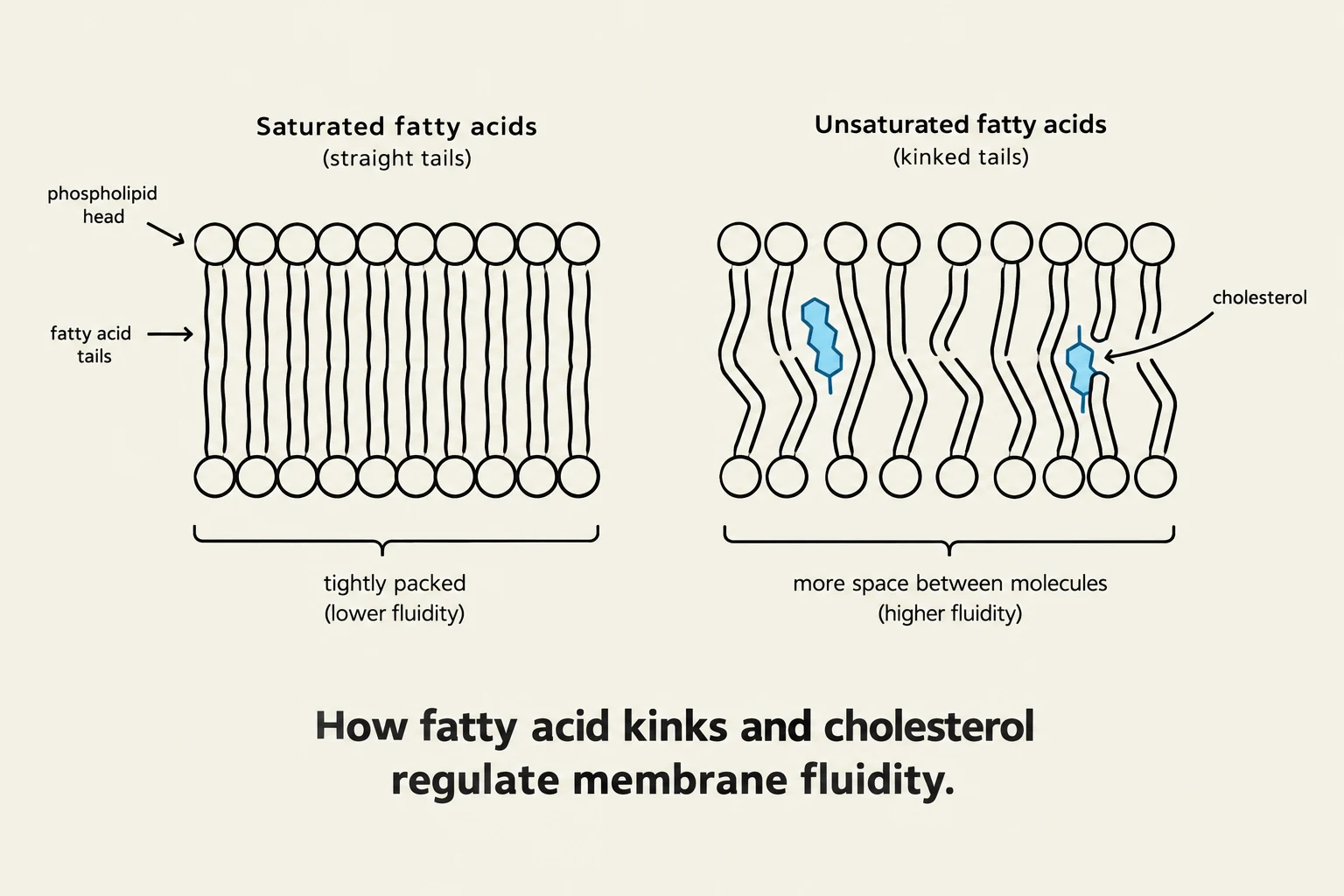
This diagram illustrates how saturated and unsaturated fatty acids, along with cholesterol, affect the fluidity of cell membranes. Saturated fatty acids with straight tails pack tightly, resulting in lower fluidity, while unsaturated fatty acids with kinked tails create more space between molecules, leading to higher fluidity. Cholesterol's presence further influences this balance.
The cell regulates how "liquid" or "solid" its membrane is through two primary chemical factors:
Fatty Acid Tails: The lipids in the bilayer have tails that can be saturated or unsaturated. The presence of double bonds in these fatty acid tails creates "kinks". These kinks prevent the lipid molecules from packing tightly together, which increases fluidity.
Cholesterol: The amount of cholesterol present within the membrane also acts as a regulator of fluidity, helping to stabilize the membrane across varying conditions.
Keep reading for free.
Create an account to finish this reading and make it part of your study routine.