An overview of blood's vital functions, including transport, regulation, and protection. It details the composition of blood, separating it into plasma and formed elements like erythrocytes, leukocytes, and platelets, and explains their respective roles.
Blood is a unique, fluid connective tissue that serves as the body's primary transport system. Unlike other connective tissues that consist of cells embedded in a solid or gel-like matrix, blood consists of living formed elements suspended in a nonliving liquid matrix called plasma. Most of these blood cells originate from hematopoietic stem cells located in the bone marrow.
Vital Functions of Blood
Blood is essential for maintaining homeostasis through several critical physiological roles:
Transport: It delivers oxygen (O2) and nutrients (such as glucose and amino acids) to body cells, while transporting metabolic wastes (like urea and carbon dioxide) to the lungs and kidneys for elimination. It also carries hormones from endocrine organs to their target tissues.
Regulation: Blood helps regulate body temperature by absorbing and distributing heat. It also plays a key role in maintaining osmotic pressure, which is vital for water balance in the blood and tissues.
Protection: It prevents hemorrhage through clot formation. Additionally, leukocytes (white blood cells) provide a defense mechanism against pathogens and disease.
Temperature regulation is achieved through the control of blood flow. Through vasodilation, blood vessels widen to increase blood flow to the skin, facilitating heat loss across the epidermis. Conversely, vasoconstriction narrows the vessels to conserve heat.
This diagram illustrates how blood vessel dilation and constriction affect heat loss across the epidermis, influencing body temperature.
Composition and Centrifugation
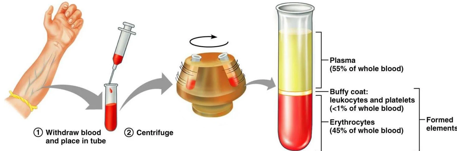
When a blood sample is centrifuged (spun at high speeds), it separates into three distinct layers based on density:
Plasma (55%): The uppermost, liquid component.
Buffy Coat (<1%): A thin, whitish layer located between the plasma and the red blood cells, containing leukocytes and platelets.
Erythrocytes (45%): The bottom layer consisting of red blood cells. The percentage of total blood volume occupied by these cells is known as the hematocrit.
Diagram illustrating the process of centrifuging blood to separate its components: plasma, buffy coat (leukocytes and platelets), and erythrocytes.
Exam Tip: Normal hematocrit values differ by biological sex. In males, it is approximately 47%±5%, while in females, it is approximately 42%±5%.
Plasma Composition
Plasma is approximately 90% water, which serves as a dissolving and suspending medium for solutes and helps absorb heat. The remaining 10% consists of several important components:
Plasma Proteins:
Albumin: The most abundant plasma protein (60%), produced by the liver; it is the primary contributor to osmotic pressure.
Globulins: These include alpha and beta globulins (transport proteins for lipids and vitamins) and gamma globulins (antibodies released by plasma cells).
Fibrinogen: A protein produced by the liver that is essential for forming the fibrin threads required for clot formation.
Solutes: These include electrolytes (such as Na+, $K^+$, $Cl^-$, and HCO3−) used to maintain pH and osmotic pressure, organic nutrients (glucose, amino acids, lipids), respiratory gases (O2 and CO2), and hormones.
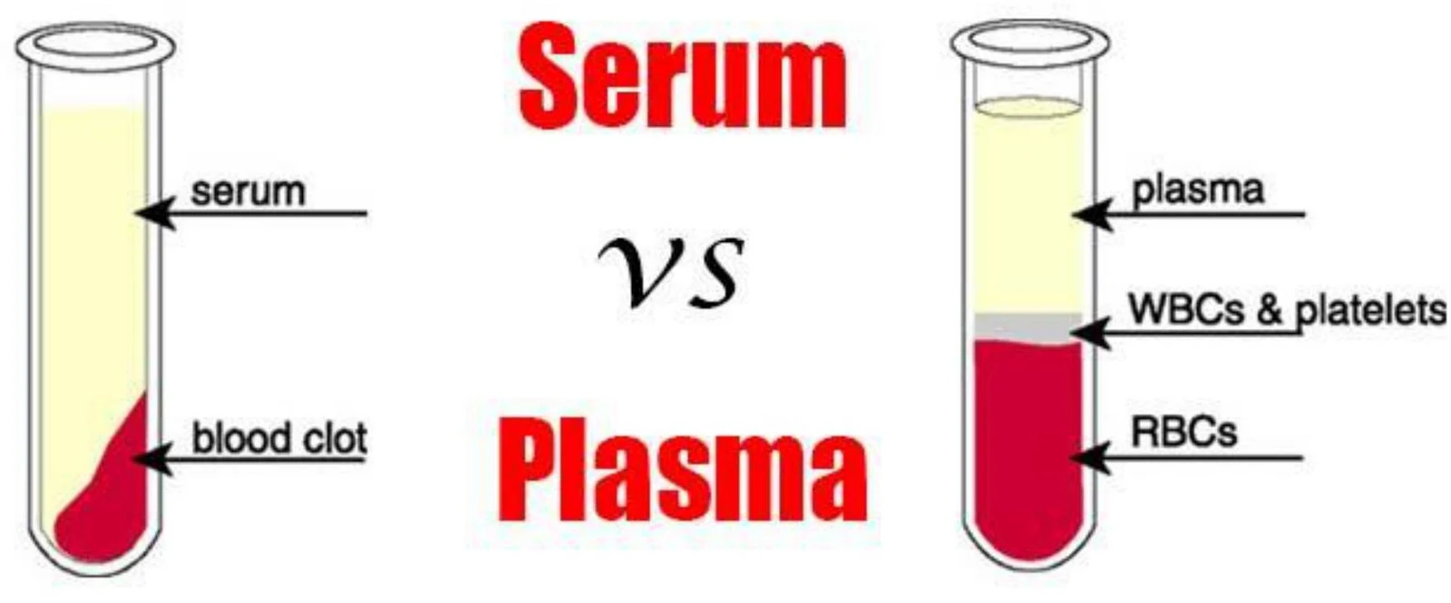
Note:Serum is defined as plasma that has had its clotting factors removed.
This illustration compares serum and plasma, highlighting their different components after blood separation in test tubes.
The Formed Elements
The formed elements are the living cells and cell fragments suspended in the plasma.
Erythrocytes (Red Blood Cells)
Erythrocytes are specialized for respiratory gas transport. They are characterized by a unique biconcave disc shape, which provides a massive surface area relative to volume for efficient gas exchange. To maximize efficiency, they are anucleate (lacking a nucleus) and contain no other organelles.
A diagram and scanning electron micrograph show the biconcave disc shape of red blood cells, with dimensions indicated.
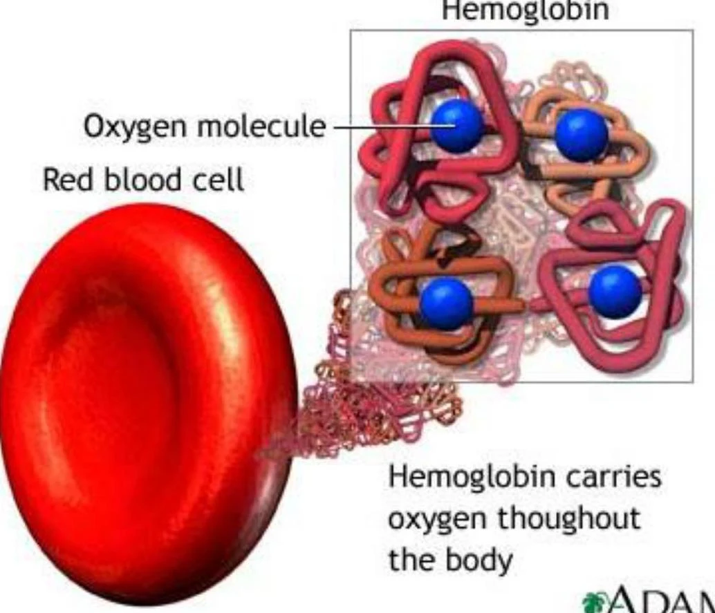
The primary functional component of an erythrocyte is hemoglobin (Hb), which makes up about 97% of the cell's volume. Hemoglobin consists of a protein called globin and a red pigment called heme, which contains an iron atom capable of binding oxygen.
Oxyhemoglobin: Hemoglobin bound to oxygen (reversible).
Carbaminohemoglobin: Hemoglobin bound to carbon dioxide (reversible).
Carboxyhemoglobin: Hemoglobin bound to carbon monoxide (irreversible and dangerous).
This illustration shows a red blood cell and hemoglobin molecules, highlighting how hemoglobin carries oxygen throughout the body.
Leukocytes (White Blood Cells)
Leukocytes are the only "complete" cells among the formed elements, meaning they possess nuclei and organelles. They are responsible for the body's immune defense and can leave the bloodstream to enter tissues via a process called diapedesis.
Leukocytes are categorized into two groups:
1. Granulocytes (contain visible granules)
Neutrophils: The most numerous WBC (50–70%). They are polymorphonuclear (having 3–6 lobes) and specialize in phagocytizing bacteria.
Eosinophils: (2–4% of WBCs) Feature a bilobed nucleus and red granules; they fight parasitic worms and are involved in allergic reactions.
Basophils: (0.5–1% of WBCs) Feature large purplish-black granules containing heparin (an anticoagulant) and histamine (a vasodilator).
2. Agranulocytes (lack visible granules)
Lymphocytes: (25% of WBCs) Crucial to immunity. They include T cells (attack virus-infected/tumor cells) and B cells (produce antibodies).
Monocytes: (3–8% of WBCs) The largest leukocytes. They leave circulation to become macrophages in the tissues, where they perform active phagocytosis.
Platelets (Thrombocytes)
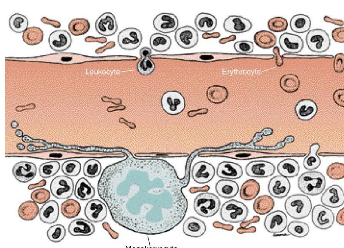
Platelets are not whole cells but rather small, discoid cytoplasmic fragments derived from much larger cells called megakaryocytes. They are essential for clot formation (hemostasis). When a blood vessel is damaged, platelets form a temporary "platelet plug" to seal the break.
Diagram illustrating leukocytes and erythrocytes within a blood vessel, alongside a megakaryocyte and other blood cells in the surrounding tissue.
Keep reading for free.
Create an account to finish this reading and make it part of your study routine.