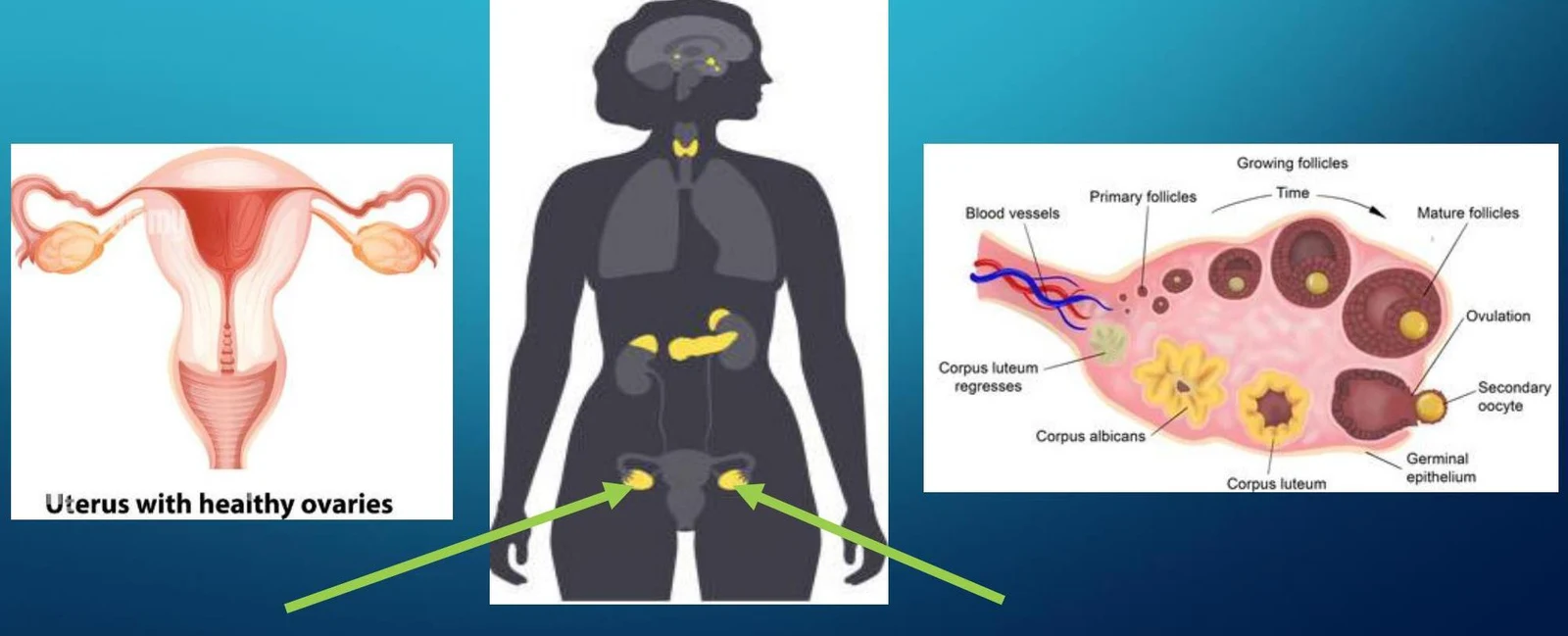
The female reproductive system is governed by a sophisticated interplay of anatomical structures and endocrine signals. Central to this system are the ovaries, paired organs located internally within the posterior pelvic fossa. Unlike the testes, which are located externally in the scrotum to maintain a temperature approximately 2∘C below core body temperature, the ovaries function within the stable internal environment of the pelvis. The primary role of the ovaries is dual: they facilitate the maturation and release of oocytes (eggs) and serve as the principal site for the production of sex steroids that regulate the menstrual cycle and secondary sexual characteristics.
The Hypothalamic-Pituitary-Ovarian (HPO) Axis
The regulation of ovarian function is not autonomous; rather, it is controlled by the Hypothalamic-Pituitary-Ovarian (HPO) axis, a complex feedback loop involving the brain and the gonads.
The process begins in the hypothalamus, which secretes Gonadotropin-Releasing Hormone (GnRH). This hormone travels to the anterior pituitary gland, stimulating it to produce and release two critical gonadotropins: Follicle-Stimulating Hormone (FSH) and Luteinizing Hormone (LH). These hormones enter the bloodstream and travel to the ovaries to initiate follicular development and ovulation.
Diagrams illustrating the female reproductive system, including the uterus and ovaries, and the process of follicular development within the ovary.
The system operates via both positive and negative feedback loops. For example, during certain stages of the cycle, rising levels of estrogen can trigger a surge in LH, whereas at other times, hormones act to inhibit the production of FSH and LH to maintain stability.
Exam Tip: In cases of primary ovarian failure (hypergonadotropic hypogonadism), you will see low estrogen levels but elevated FSH and LH levels, because the pituitary is attempting to compensate for the lack of ovarian response.
Ovarian Hormones: Estrogens, Progesterone, and Androgens
The ovaries produce several key steroid hormones that dictate the physiological changes seen throughout the menstrual cycle and throughout life.
Estrogens
Primarily in the form of estradiol, estrogens are responsible for the development of secondary female sex characteristics, including the growth of breasts, the vagina, and the uterus. Estrogens also play a vital role in systemic health, promoting the maintenance of skin, blood vessels, bone density, and central nervous system function. A significant decline in estrogen is the hallmark of menopause, which often leads to ovarian atrophy.
Progesterone
Progesterone levels rise significantly following ovulation. Its primary function is to prepare the endometrium (the uterine lining) for potential embryo implantation by inducing endometrial gland secretions.
If pregnancy occurs: The placenta takes over progesterone production, keeping levels high to prevent menstruation.
If no pregnancy occurs: Progesterone levels fall, triggering the shedding of the endometrial lining (menstruation).
Androgens
While often associated with male biology, androgens (primarily testosterone) are also produced by the ovaries. They contribute to a woman's libido, sense of well-being, and muscle/bone health. However, in excess—often seen in conditions like Polycystic Ovary Syndrome (PCOS)—androgens can cause hirsutism (excessive hair growth) and the development of male-pattern secondary sexual features.
The Menstrual Cycle: Follicular and Luteal Phases
The ovarian cycle is divided into two distinct functional phases, separated by the event of ovulation.
The Follicular Phase
This phase begins on the first day of menses and continues until ovulation.
Early Phase: Estrogen and progesterone levels are initially very low.
Growth: An increase in FSH stimulates the growth of ovarian follicles.
Estrogen Rise: As these follicles develop, they secrete increasing amounts of estrogen. This estrogen stimulates the uterine epithelial cells and blood vessels to thicken the endometrium.
Peak: Estrogen levels peak approximately one day before ovulation.
The LH Surge and Ovulation
The peak in estrogen concentration triggers a dramatic spike in Luteinizing Hormone, known as the LH surge. This surge is the critical signal that culminates in ovulation—the extrusion of the ovum from the follicle.
The Luteal Phase
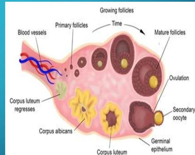
Following ovulation, the remains of the ruptured follicle transform into the corpus luteum, marking the start of the luteal phase.
Progesterone Dominance: The primary goal of this phase is the secretion of progesterone to support a potential pregnancy.
End of Cycle: If fertilization does not occur, the corpus luteum regresses, leading to a gradual decline in both progesterone and estrogen. This hormonal drop causes the loss of the endometrial blood supply, resulting in the shedding of the lining (menses) approximately 14 days after ovulation.
Diagram illustrating the stages of follicular development and ovulation within the ovary.
Clinical Correlations and Development
Tanner Staging
The onset of puberty is marked by the activation of the HPO axis, leading to increased FSH and LH levels. Clinicians use Tanner Staging to track the progression of secondary sexual characteristics in both males and females. In females, this includes the development of breast tissue (from prepubertal stages to a mature stage where the areola recedes into the breast) and the progression of pubic hair from fine lanugo-type hair to thick, terminal hair that eventually spreads to the inner thighs.
Clinical Abnormalities
Disruptions in the HPO axis or ovarian function can lead to significant clinical issues:
Amenorrhea: The absence of menses. This can be primary (never menstruating by age 16) or secondary (loss of menses for 3–6 months). Causes range from hypothalamic issues (stress, diet, weight loss) to pituitary tumors or ovarian failure.
Polycystic Ovary Syndrome (PCOS): A common disorder characterized by chronic anovulation, infertility, and hirsutism. Ultrasound typically reveals multiple ovarian cysts.
Infertility: Can result from various targets, including decreased GnRH from the hypothalamus, organ failure in the ovaries, or inadequate progesterone output preventing endometrial support.
Clinical Note: When evaluating a patient for secondary amenorrhea, clinicians often follow a diagnostic pathway starting with a β-hCG test to rule out pregnancy, followed by specific hormone assays to differentiate between hypothalamic, pituitary, or ovarian causes.
Keep reading for free.
Create an account to finish this reading and make it part of your study routine.