Foundations of Obstetrics and Gynecology: From Embryology to Clinical Practice
An integrated overview of female reproductive health, covering pelvic anatomy and embryogenesis, the Hypothalamic-Pituitary-Ovarian (HPO) axis, and clinical practices in cervical and breast health.
Understanding female reproductive health requires a dual mastery of developmental biology and clinical application. To treat conditions ranging from menstrual irregularities to cervical neoplasia, a clinician must first grasp the intricate anatomical scaffolding of the pelvis and the hormonal orchestration of the HPO axis. This article integrates the embryological origins of these structures with the clinical frameworks used in modern obstetric and gynecological practice.
Anatomy and Embryogenesis of the Female Pelvis
The female pelvis is a complex arrangement of ligaments, vessels, and organs that must provide both stability and mobility. The uterus is positioned between the bladder and the rectum. Its stability is maintained by several key ligamentous suspensions: the uterosacral ligaments connect the uterus to the sacrum, while the cardinal ligaments attach to the sides of the uterus inferior to the broad ligament. The round ligament extends from the uterus to the labia majora.
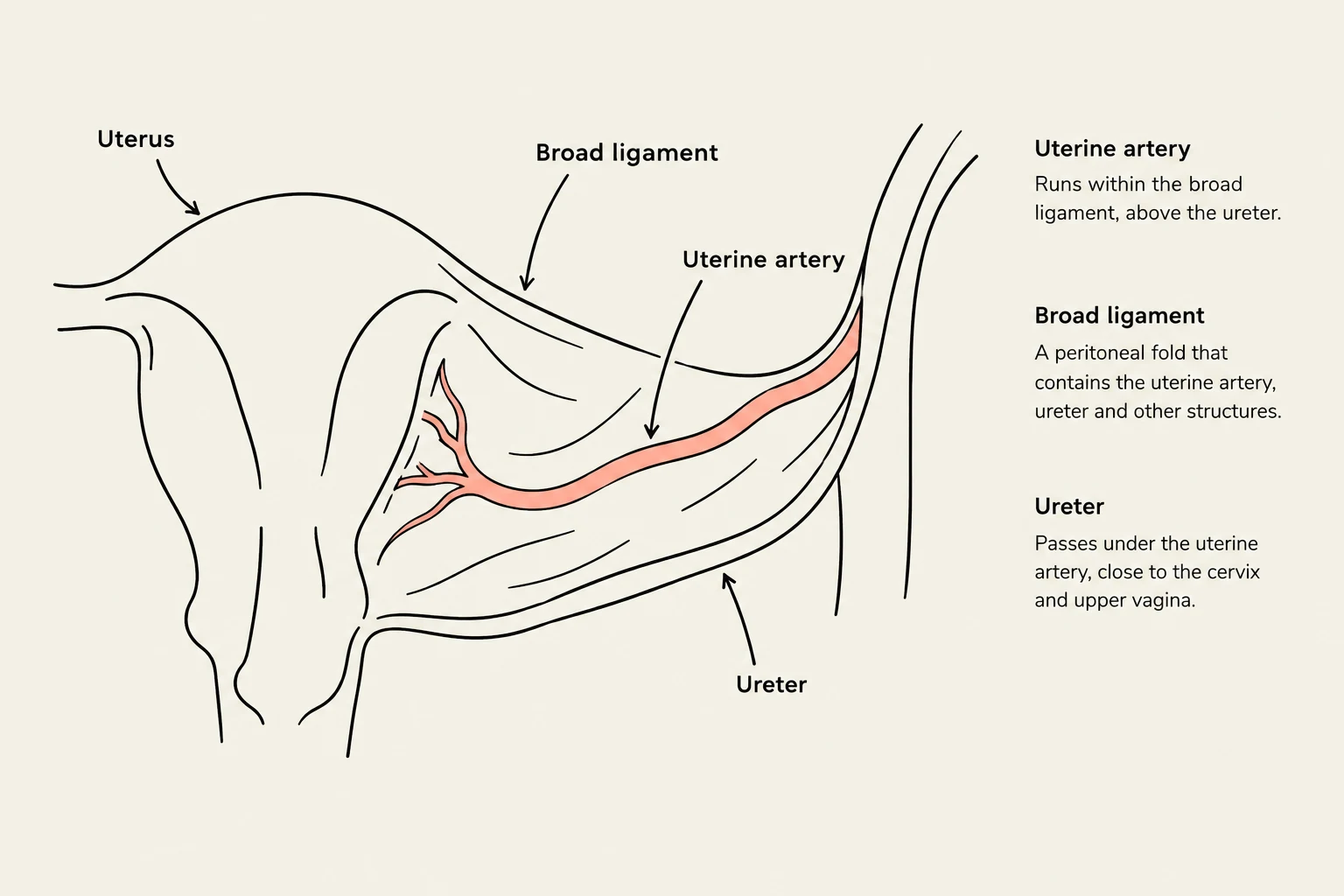
The ovaries are suspended by the infundibulopelvic (suspensory) ligament, which is a critical structure because it contains the ovarian vessels. The broad ligament serves as a wide fold of peritoneum overlying the uterus and adjacent structures, housing the uterine arteries, veins, and ureters.
Clinical Pearl: The "Water Under the Bridge" Rule
In pelvic surgery, clinicians must be acutely aware of the relationship between the ureters and the uterine arteries. The ureters run inferior to (below) the uterine arteries. Accidental ligation of the ureter during a hysterectomy is a significant surgical complication.
An illustration showing the anatomical relationship between the uterus, broad ligament, uterine artery, and ureter.
Vascular supply to these organs is primarily derived from the branches of the internal iliac arteries. The anterior division provides the umbilical, uterine, superior vesical, vaginal, obturator, middle rectal, internal pudendal, and inferior gluteal arteries, while the posterior division supplies the iliolumbar, lateral sacral, and superior gluteal arteries.
Developmental Origins
The structures of the pelvis are the result of precise embryological movements. The ovary begins development in the early fetal period when mesothelial cells form cortical cords that invade the medulla. By week 16, these cords surround primordial germ cells, which differentiate into oogonia. These cells undergo mitosis and enter the first meiotic division to become primary oocytes. When a primary oocyte is surrounded by squamous follicular cells, it forms a primordial follicle.
The external genitalia, or vulva, develop from the cloacal fold during the 3rd week. This fold divides into the genital tubercle (the primordial phallus), the urethral folds, and the anal fold. The genital (labioscrotal) swellings subsequently differentiate into the labia in females. The vulva itself comprises the labia majora (adipose-filled folds), labia minora (narrow skin folds), the clitoris (consisting of the glans and two crura), and the vestibule.
Reproductive Physiology and the HPO Axis
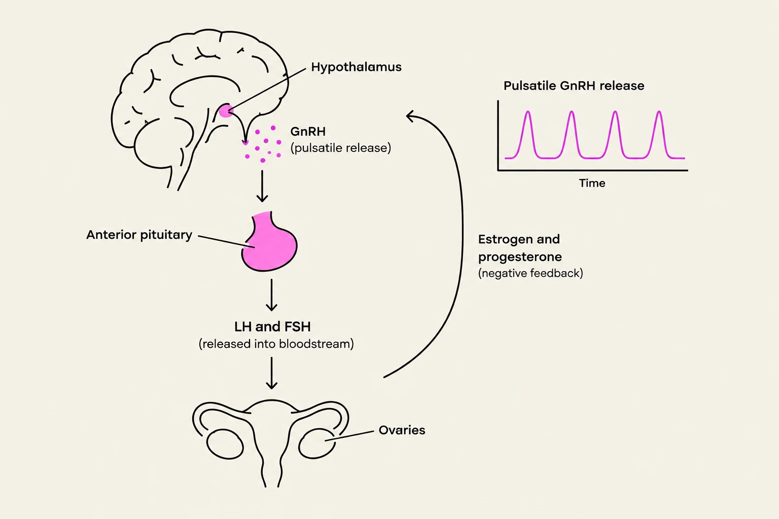
The regulation of the menstrual cycle is governed by the Hypothalamic-Pituitary-Ovarian (HPO) axis. This system utilizes complex feedback loops to coordinate the maturation of an oocyte with the preparation of the endometrium.
The cycle, which typically lasts 28 days, is divided into three distinct phases:
Menstruation and the Follicular Phase: Low levels of estrogen and progesterone trigger the release of FSH (Follicle-Stimulating Hormone). FSH stimulates follicle growth; the dominant follicle produces increasing amounts of estradiol, which drives endometrial cell proliferation.
Ovulation: As estradiol levels peak, they trigger a positive feedback loop that causes an LH (Luteinizing Hormone) surge. This surge results in the expulsion of the oocyte from the follicle. Meiosis resumes but arrests in metaphase II until fertilization occurs.
Luteal Phase: The remains of the follicle transform into the corpus luteum, which secretes high levels of progesterone. Progesterone converts the proliferative endometrium into a secretory endometrium, characterized by thickened, twisting blood vessels and tortuous glands. If conception does not occur, the corpus luteum becomes the corpus albicans (a white scar), and progesterone levels drop, triggering menstruation. If conception occurs, hCG (human chorionic gonadotropin) sustains the corpus luteum until the placenta takes over progesterone production at approximately 9–10 weeks gestation.
Diagram illustrating the hormonal regulation of the female reproductive system, showing the roles of the hypothalamus, anterior pituitary, ovaries, and hormones like GnRH, LH, FSH, estrogen, and progesterone in a feedback loop.
Hormonal Summary Table
Hormone
Primary Source
Primary Function
GnRH
Hypothalamus
Stimulates pulsatile release of FSH and LH
FSH
Anterior Pituitary
Promotes granulosa cell proliferation
LH
Anterior Pituitary
Stimulates theca cells to produce androgens
Estradiol
Ovary (Follicle)
Drives endometrial proliferation
Progesterone
Corpus Luteum
Drives endometrial differentiation (secretory)
Clinical Practice: History, Examination, and Pathology
In a clinical setting, a gynecological history must be comprehensive, covering menstrual history (menarche, LMP, frequency, duration, and flow), contraceptive use, sexual history (number of partners, history of STIs), and symptoms like dyspareunia. Clinicians must also accurately record gravidity (total number of pregnancies) and parity (number of births, categorized by term, preterm, abortion, and living).
Cervical and Breast Health
Cervical cancer screening is a cornerstone of preventive gynecology. Current guidelines recommend:
Ages 21–29: Pap smear every 3 years.
Ages 30–65: Co-testing with HPV (Human Papillomavirus) testing every 5 years.
The primary driver of cervical dysplasia and cancer is high-risk HPV (specifically types 16, 18, 31, and 33). Dysplasia is categorized histologically as Cervical Intraepithelial Neoplasia (CIN), divided into CIN 1, 2, and 3 based on the depth of cellular invasion. The HPV vaccine, which uses non-infectious virus-like particles to stimulate IgG production, is administered in a 3-dose series at 0, 2, and 6 months to prevent acquisition of these high-risk types.
Breast health evaluation involves palpation to check for masses or tenderness, followed by diagnostic imaging such as mammography, ultrasound, or MRI. Risk factors for breast cancer include BRCA 1/2 mutations, nulliparity, early menarche, and late menopause.
Exam Tip: Breast Disease Evaluation
When a patient presents with nipple discharge, note whether it is unilateral or bilateral, and whether it is spontaneous or expressed. Discharge types can range from milky and green to bloody, which significantly alters the diagnostic pathway (e.g., ductography or ductal excision).
By integrating these anatomical, physiological, and clinical principles, healthcare providers can more effectively navigate the complexities of female reproductive health.
Keep reading for free.
Create an account to finish this reading and make it part of your study routine.