An exploration of human foot development from embryonic stages to clinical assessment, covering limb formation, skeletal maturation, and common postural deviations like in-toeing.
The human foot is a masterpiece of biological engineering, transitioning from a simple embryonic bud into a complex, multi-axial lever capable of both shock absorption and powerful propulsion. Understanding foot posture requires a holistic view that spans from the molecular signaling of the womb to the biomechanical alignment of the growing child. This article explores the developmental milestones of limb formation, the physiological evolution of foot structure, and the clinical tools used to assess common postural deviations.
Embryonic Limb Development
The journey of the foot begins long before birth. During the fourth week of gestation, limb buds emerge as small protrusions composed of a core of lateral plate mesoderm covered by a layer of ectoderm. The precise shaping of these buds is governed by specialized signaling centers that dictate growth along three distinct axes.
The Apical Ectodermal Ridge (AER) is a critical structure at the distal tip of the limb bud. It utilizes FGF8 and FGF10 signaling to drive proximal-to-distal outgrowth, essentially telling the limb to grow "outward" from the body. Simultaneously, the Zone of Polarizing Activity (ZPA) manages the anterior-posterior axis (the thumb-to-pinky or big toe-to-little toe axis) through the secretion of Sonic Hedgehog (Shh).
As development progresses into weeks 5 and 6, the limb plates transform into digital rays. A pivotal moment occurs during week 7, when the limbs undergo rotation. While the upper limbs rotate 90° laterally, the lower limbs rotate 90° medially, a movement that positions the big toe on the medial aspect of the foot. By week 8, the individual digits are finalized through apoptosis, the programmed cell death of the interdigital webbing.
Clinical Pearl: The period between weeks 4 and 8 is the "critical window" for limb development. Disruptions in signaling during this time can lead to permanent malformations such as syndactyly (fused digits), polydactyly (extra digits), or clubfoot.
Diagram illustrating the proximal-distal outgrowth and anterior-posterior patterning of a limb bud, and how these axes work together during development.
Developmental Factors in Foot Growth
As a child grows, the foot undergoes significant morphological and skeletal changes. An infant's foot often appears flat; this is not necessarily due to a lack of arches, but rather the presence of thick subcutaneous fat padding. The development of the medial longitudinal arch is a gradual process that typically becomes evident between ages 6 and 10.
The skeletal framework matures through a predictable pattern of ossification:
At Birth: The talus and calcaneus are already ossified.
Ages 2–3: The navicular begins to ossify.
Ages 3–5: The remaining tarsal bones undergo ossification.
Adolescence: Growth plates generally close between ages 14 and 18.
Growth rates are most rapid in the first two years of life, with the foot reaching approximately 90% of its adult length by age 12 in girls and age 14 in boys.
Clinical Assessment: The Foot Posture Index (FPI-6)
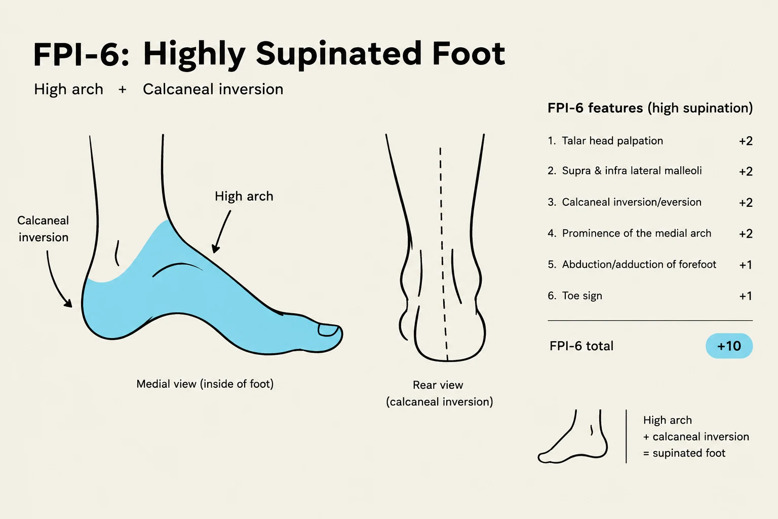
In clinical practice, assessing the static posture of the foot is essential for identifying biomechanical risks. The Foot Posture Index (FPI-6) is a validated tool used to score the alignment of the foot on a scale ranging from −12 to +12.
The assessment is divided into two main anatomical regions:
Rearfoot Criteria: Includes the palpation of the talar head, the curves above and below the lateral malleolus, and the calcaneal position in the frontal plane.
Forefoot Criteria: Includes bulging at the talonavicular joint, the congruence of the medial arch, and the abduction or adduction of the forefoot relative to the rearfoot.
The total score allows clinicians to categorize the foot posture as follows:
Total Score
Classification
−12 to −5
Highly Supinated
−4 to −1
Supinated
0 to +5
Normal
+6 to +9
Pronated
+10 to +12
Highly Pronated
This illustration depicts the FPI-6 (Foot Posture Index) assessment for a highly supinated foot, characterized by a high arch and calcaneal inversion. It includes visual representations of the medial and rear views of the foot, along with a list of features and their corresponding scores that contribute to the FPI-6 total.
Biomechanics of In-Toeing
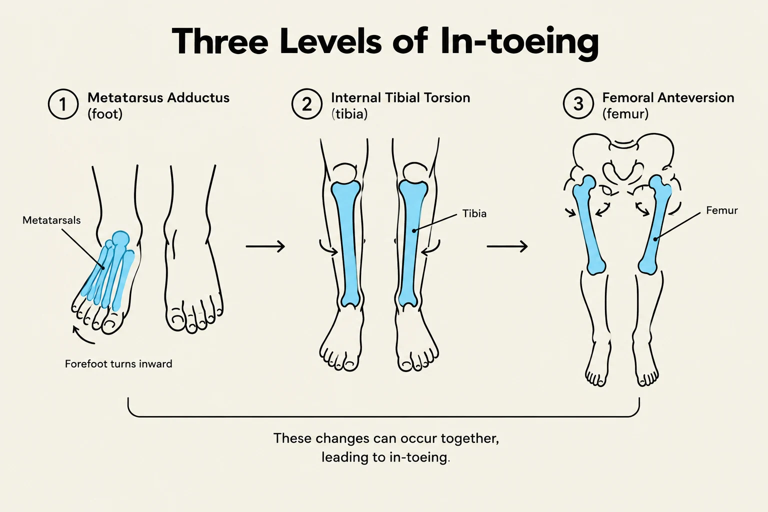
One of the most common presentations in pediatric orthopedics is in-toeing, characterized by a negative foot progression angle (where the normal range is roughly −3∘ to +20∘). To effectively treat in-toeing, a clinician must identify whether the deviation originates at the foot, the tibia, or the femur.
The Three Levels of Deviation
Foot Level (Metatarsus Adductus): Most common in infancy. The forefoot is turned inward relative to the hindfoot, often creating a "bean-shaped" appearance of the lateral foot border.
Tibia Level (Internal Tibial Torsion): Typically observed in children aged 1–3 years. This is characterized by a negative thigh-foot angle.
Femur Level (Femoral Anteversion): Common in children aged 3–8 years. This involves excess hip internal rotation and is often associated with "W-sitting" postures.
Diagram illustrating the three levels of in-toeing: Metatarsus Adductus (foot), Internal Tibial Torsion (tibia), and Femoral Anteversion (femur). These conditions can occur together and lead to in-toeing.
Exam Tip: When assessing in-toeing, do not just look at the feet. You must perform a full rotational profile, including the foot progression angle, thigh-foot angle, and the transmalleolar axis.
While many cases of in-toeing resolve spontaneously as the child grows and the bones "derotate," surgery is a last resort. It is generally reserved for severe, persistent cases that do not improve after age 8–10.
Keep reading for free.
Create an account to finish this reading and make it part of your study routine.