Female Reproductive System: Anatomy and Physiology
An in-depth explanation of the female reproductive system, covering its functional anatomy, oogenesis, the ovarian and uterine cycles, hormonal control by oestrogen and progesterone, and related clinical terms.
The female reproductive system is a complex network of organs designed for the production of gametes, the facilitation of fertilization, and the support of a developing fetus. It involves a highly coordinated interplay between anatomical structures and endocrine signaling, primarily through the hormones oestrogen and progesterone.
Functional Anatomy of the Reproductive Organs
The female reproductive system is divided into primary sex organs (the gonads), accessory ducts, and external genitalia.
Internal Organs
Ovaries: The paired primary sex organs (gonads) located on either side of the uterus. They are responsible for oogenesis and the secretion of reproductive hormones.
Fallopian Tubes (Uterine Tubes): These tubes extend from the uterus toward the ovaries. The end near the ovary expands into the ampulla and terminates in a funnel-shaped, ciliated infundibulum with finger-like fimbriae. The fimbriae create currents to carry the ovulated oocyte into the tube, which serves as the site for fertilization.
Uterus: A hollow, thick-walled organ located in the pelvis, positioned anterior to the rectum and posterosuperior to the bladder. It consists of the fundus (superior region), the body, the isthmus (narrowed region), and the cervix (the neck projecting into the vagina).
Vagina: A thin-walled tube extending from the cervix to the exterior of the body, serving as the passageway for birth, menstrual flow, and copulation.
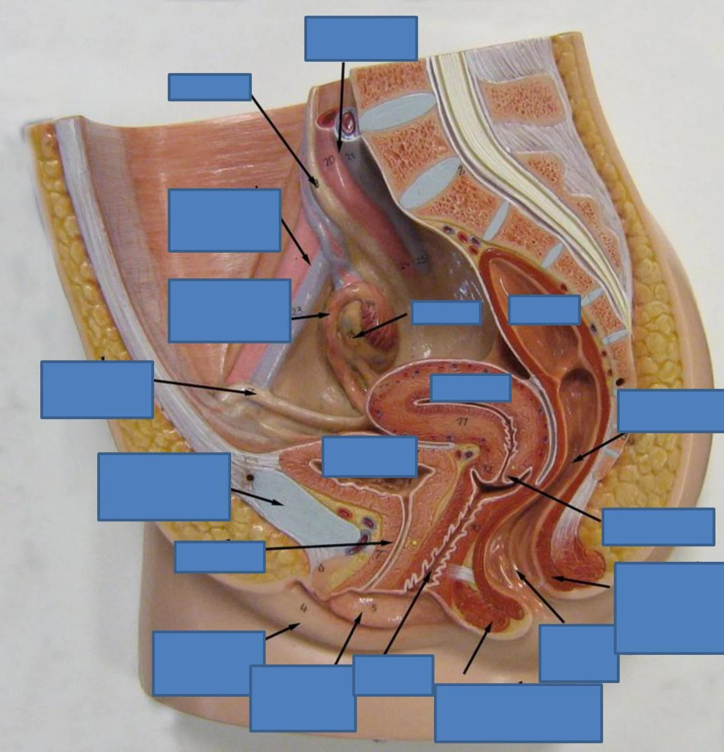
Anatomical model of the female pelvic organs, including the uterus, ovaries, and surrounding structures.
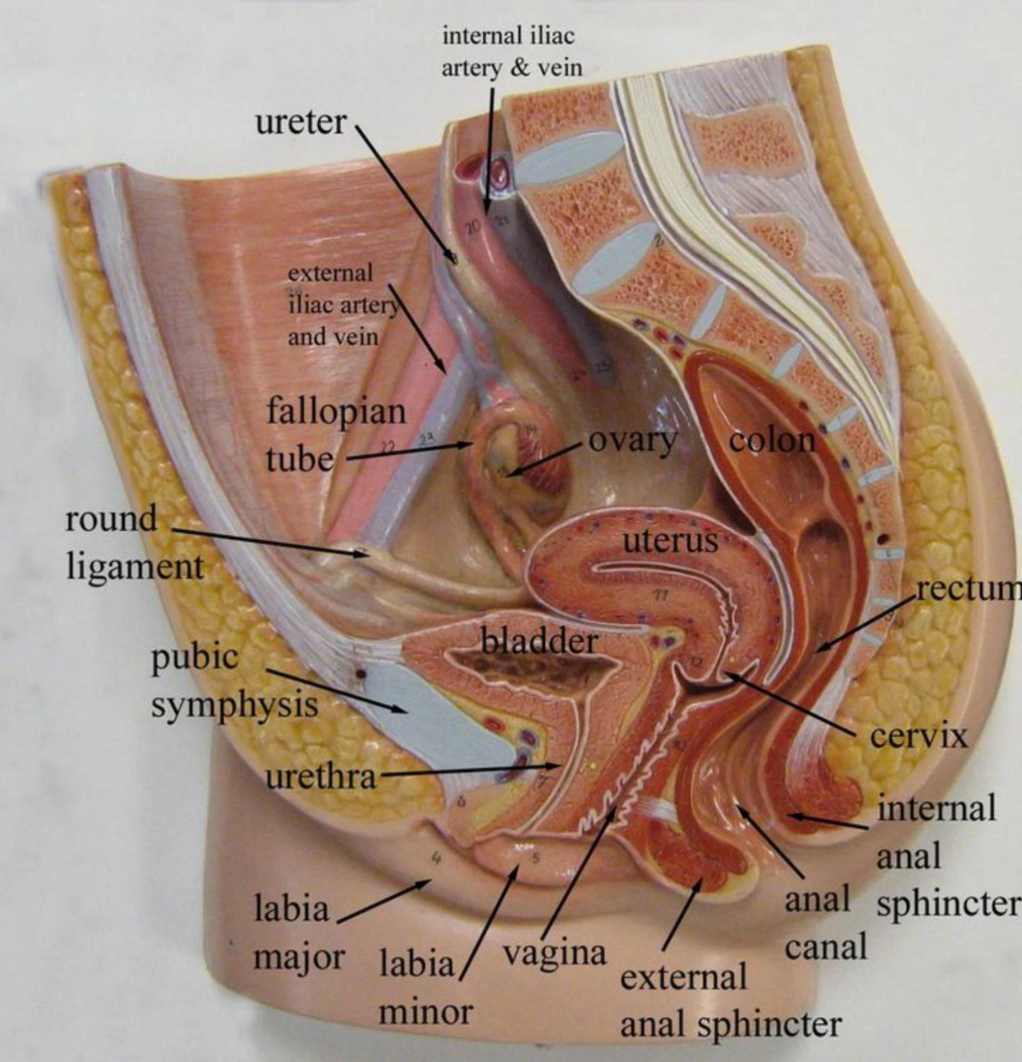
Anatomical model of the female pelvic organs, including the uterus, bladder, rectum, ovaries, fallopian tubes, ureters, and associated blood vessels and ligaments.
Uterine Support and Layers
The uterus is held in position by several key ligaments, including the broad ligament (lateral support), cardinal ligaments, lateral cervical ligaments, sacro-uterine ligaments, and the round ligament (which binds the anterior uterine wall to the labia majora).
Diagram of the female reproductive system, illustrating the uterus, fallopian tubes, ovaries, cervix, and vagina, along with supporting ligaments.
The uterine wall is composed of three distinct layers:
Perimetrium: The outermost serous layer.
Myometrium: The middle layer of smooth muscle.
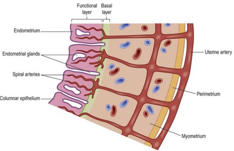
Endometrium: The mucosal lining. This is divided into the stratum functionalis (the innermost layer that undergoes cyclic changes and is shed during menstruation) and the stratum basalis (the outer layer that regenerates the functionalis).
Diagram illustrating the layers of the uterine wall, including the endometrium with its functional and basal layers, endometrial glands, spiral arteries, columnar epithelium, uterine artery, perimetrium, and myometrium.
External Genitalia and Mammary Glands
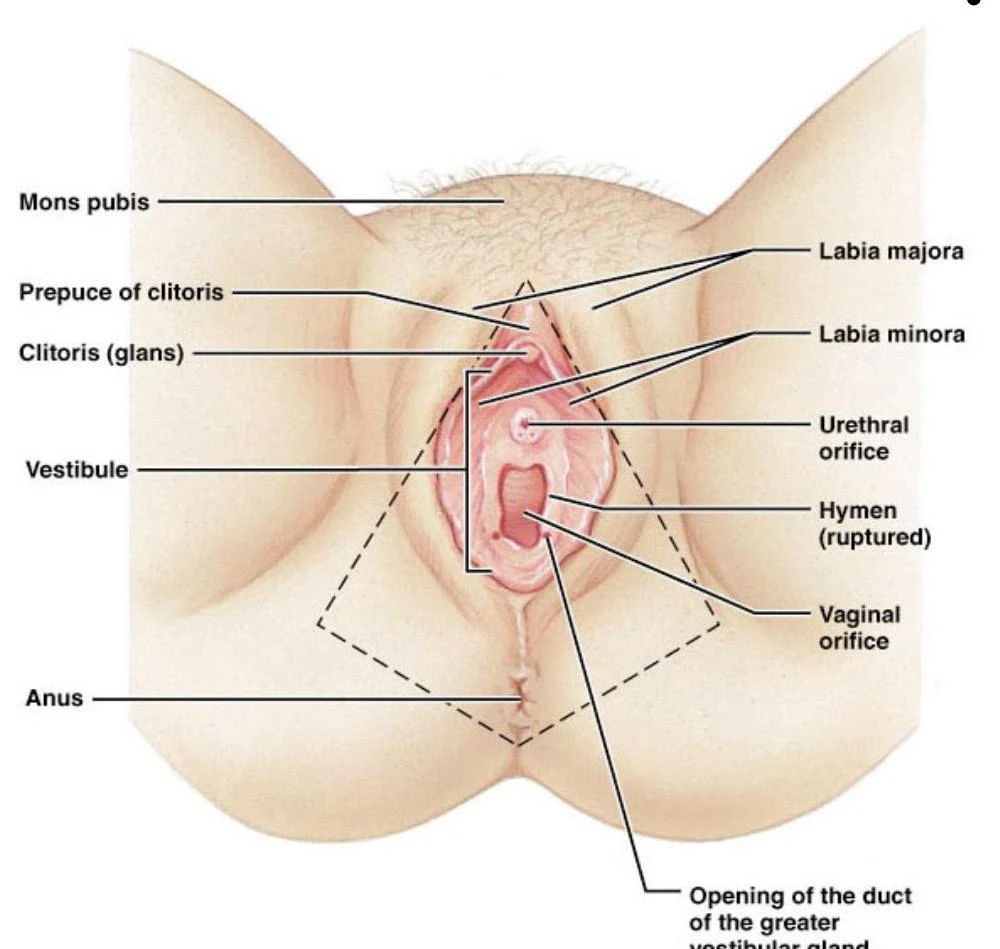
The external genitalia, collectively known as the vulva, include the mons pubis, labia majora, labia minora, and the clitoris. Additionally, the mammary glands are modified sweat glands consisting of 15–25 lobes that produce milk during lactation.
Anatomical illustration of the female external genitalia, labeling the mons pubis, labia majora and minora, clitoris, vestibule, urethral orifice, hymen, vaginal orifice, and anus.
Oogenesis and the Ovarian Cycle
Oogenesis is the process of producing a female gamete (ovum) via meiosis. This process begins before birth, where oogonia divide to produce primary oocytes that remain arrested in prophase I until puberty. At puberty, a primary oocyte is activated each month, completing meiosis I to form a secondary oocyte and a first polar body. Meiosis II is only completed if the secondary oocyte is penetrated by a sperm.
Diagram illustrating oogenesis and follicle development in the ovary, showing the stages from oogonium to ovulated secondary oocyte, including meiotic events and hormonal influences.
The ovarian cycle describes the monthly maturation of the ovum and is divided into three phases:
Follicular Phase (Days 1–14): Regulated by FSH and LH, primordial follicles grow into primary, secondary, and finally Graafian follicles.
Ovulation (Midcycle): The ovary wall ruptures, expelling the secondary oocyte.
Luteal Phase (Days 14–28): The ruptured follicle becomes the corpus luteum, which secretes progesterone and oestrogen. If no pregnancy occurs, it degenerates into the corpus albicans.
Diagram of the ovarian cycle showing follicular development, ovulation, and corpus luteum formation.
The Uterine (Menstrual) Cycle
The uterine cycle consists of cyclic changes in the endometrium in response to ovarian hormones.
Menstrual Phase (Days 1–5): If fertilization does not occur, progesterone levels fall, causing the spiral arteries to constrict and the stratum functionalis to shed.
Proliferative Phase (Days 6–14): The endometrium rebuilds itself under the influence of oestrogen.
Secretory Phase (Days 15–28): The endometrium prepares for potential embryo implantation under the influence of progesterone, which converts the lining into a secretory mucosa.
This diagram illustrates the phases of the ovarian and uterine cycles, along with the corresponding changes in pituitary and ovarian hormone levels throughout the menstrual cycle.
Hormonal Control and Physiological Effects
Oestrogen and Progesterone
These hormones are central to female reproductive health and the development of secondary sex characteristics.
Feature
Oestrogen Effects
Progesterone Effects
Feedback
Negative feedback on FSH/LH; Positive feedback induces LH surge for ovulation
Breast development; widening of pelvis; pubertal growth spurt
Breast development; sustains pregnancy
Other
Increases prolactin secretion
Increases body temperature; inhibits T-lymphocytes to tolerate fetus
Contraceptive Pills
Oral contraceptives (the pill) use synthetic versions of these hormones (oestrogen and progestin) to prevent pregnancy. They work by:
Preventing ovulation.
Thickening cervical mucus to block sperm entry.
Thinning the endometrium to prevent implantation.
Clinical Terms and Sexual Response
Clinical Terminology
Menarche: The onset of menstruation.
Menopause: The cessation of the menstrual cycle.
Amenorrhoea: The absence of menstruation.
Dysmenorrhoea: Painful menstruation.
Female Sexual Response
The female sexual response involves engorgement of the clitoris, vaginal mucosa, and breasts with blood. Orgasm is characterized by muscle tension, increased pulse/blood pressure, and rhythmical contractions of the uterus.
Exam Tip: Remember that the stratum functionalis is the part of the endometrium that is shed during the menstrual phase due to the degeneration of spiral arteries, while the stratum basalis remains to rebuild the lining.
Keep reading for free.
Create an account to finish this reading and make it part of your study routine.