


Understanding the female pelvis requires a dual focus on its complex structural architecture and the precise embryological processes that form these organs. For clinicians, particularly in obstetrics and gynecology, mastering the spatial relationships between ligaments, vessels, and the urinary tract is essential for safe surgical intervention and the diagnosis of pelvic pathologies.
Ligamentous Suspensions and Spatial Relationships
The uterus and ovaries are not floating freely within the pelvic cavity; they are held in place by a sophisticated network of ligaments that provide both structural support and a pathway for vital neurovascular structures.
Key Ligaments of the Uterus and Ovaries
- Infundibulopelvic (Suspensory) Ligament: This ligament connects the ovaries to the posterior abdominal wall. Crucially, it serves as the conduit for the ovarian vessels.
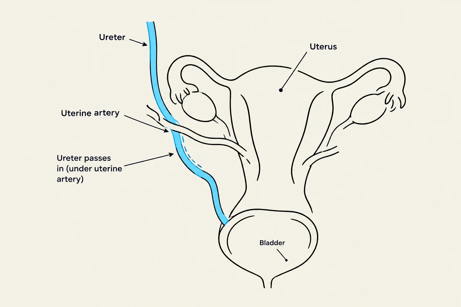
- Broad Ligament: A wide fold of peritoneum that overlies the structures adjacent to the uterus. It contains the uterine arteries, uterine veins, and the ureters.
- Uterosacral Ligament: This provides posterior support by connecting the uterus to the sacrum.
- Cardinal Ligament: Located at the side of the uterus, inferior to the broad ligament, this structure contains the uterine artery.
- Round Ligament: This ligament attaches the uterus to the labia majora, helping to maintain the anteverted position of the uterus.
Clinical Pearl: The "Water Under the Bridge" Rule In pelvic surgery, a critical anatomical relationship must be respected: the ureters run inferior to (below) the uterine arteries. Misidentifying this relationship during procedures like a hysterectomy can lead to accidental ureteral injury.
Keep reading for free.
Create an account to finish this reading and make it part of your study routine.
Continue reading freeAlready have an account? Log in
Updated