This content provides a comprehensive overview of the human eye's anatomy, methods for conducting focused eye assessments (both subjective and objective), and discusses common vision changes that occur throughout a person's lifespan, from infancy to old age.
The eyes are complex sensory organs responsible for processing visual information. Because vision is critical for independence, safety, and quality of life, healthcare providers must be proficient in performing both subjective and objective assessments. A focused eye assessment allows clinicians to identify normal physiological function, detect early signs of pathology, and monitor changes across a patient's lifespan.
Subjective Data Collection
The assessment begins with a thorough interview. Subjective data consists of what the patient reports, such as changes in vision, pain, or discomfort. When a patient presents with a specific ocular symptom—such as blurred vision, sudden loss of sight, or eye pain—clinicians should use a structured mnemonic to ensure no critical information is missed.
The PQRST(A)U Guide
To systematically explore a symptom, use the PQRST(A)U mnemonic:
P (Provocation/Palliation): What makes the symptom better or worse? (e.g., Does reading or bright light affect the pain?)
Q (Quality/Quantity): How would you describe the sensation? (e.g., Is the pain sharp, burning, or aching? Is the vision blurry or doubled?)
R (Region/Radiation): Which eye is affected, and does the sensation spread?
S (Severity): How intense is the symptom on a scale of 0–10?
T (Timing): When did it start? Was the onset sudden or gradual? Is it constant or intermittent?
A (Associated symptoms): Are there other issues like redness, swelling, headache, nausea, or light sensitivity?
U (Understanding): What does the patient believe is causing the problem?
Clinical Alert: Sudden vision loss, flashes of light, "floaters," or a sensation like a "curtain" being pulled over the vision require immediate, urgent evaluation.
Objective Data Collection
Objective data is gathered through physical inspection and specialized testing. This process moves from assessing how the patient sees (acuity and fields) to how the eyes move (muscles) and finally to the physical structures of the eye itself.
Visual Acuity and Fields
Central Visual Acuity
The first step is testing how clearly a patient sees.
Distance Vision: This is assessed using the Snellen Chart, which consists of lines of letters in decreasing sizes. The patient stands approximately 6 meters (20 feet) from the chart. Results are recorded as a fraction, such as 20/20.
Near Vision: This is assessed using a handheld vision screener with various-sized letters, held approximately 35 cm (14 inches) from the patient.
Peripheral Vision
The Confrontation Test is used to measure the patient's peripheral (side) vision. The examiner sits at eye level with the patient. Both the examiner and the patient cover one eye, and the examiner moves a finger from the periphery toward the center. The patient reports when they first see the finger. A normal finding is that the peripheral fields are intact and equal to the examiner's.
Extraocular Muscle (EOM) Function
To ensure the eyes move in a coordinated fashion, clinicians assess the six extraocular muscles. This is done through two primary methods:
Corneal Light Reflex (Hirschberg Test): Shine a penlight toward both eyes and observe the reflection of light on the corneas. In a normal finding, the light reflection appears symmetrical in both eyes.
Diagnostic Positions Test (Six Cardinal Fields of Gaze): Ask the patient to follow your finger with their eyes without moving their head. The finger is moved through six specific directions to ensure smooth, coordinated movement. A normal finding is movement without nystagmus (involuntary shaking).
External Inspection and Pupillary Response
The clinician must inspect the external structures, including the eyelids, eyelashes, conjunctiva, sclera, and cornea.
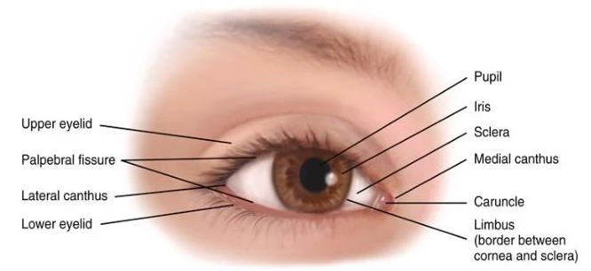
Diagram of the human eye with labels for its various parts including the pupil, iris, sclera, eyelids, and canthi.
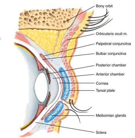
This diagram illustrates the anatomy of the human eye, labeling key structures such as the bony orbit, orbicularis oculi muscle, conjunctiva, cornea, tarsal plate, Meibomian glands, and sclera.
A critical component of the physical exam is assessing the pupillary response using the PERRLA acronym:
Pupils
Equal (Both are approximately the same size)
Round (Circular in shape)
Reactive to
Light (Constrict when exposed to light via direct and consensual reflexes)
Accommodation (Pupils constrict and eyes converge when focusing on a near object)
Note on Accommodation: To test this, have the patient look at a distant object, then move your finger about 7–8 cm from their nose and ask them to focus on it.
Lifespan Changes in Vision
Vision is a dynamic process that changes significantly from birth through old age.
Infancy and Childhood
Vision is not fully developed at birth. While the pupillary light reflex is present, eye movements may be briefly uncoordinated in newborns. As children grow, they gradually develop visual tracking and focusing abilities. During childhood, it is vital to screen for strabismus (misaligned eyes) and amblyopia (lazy eye), as early detection is crucial for proper visual development and learning.
The Aging Eye
As patients age, several physiological and pathological changes become more common:
Presbyopia: The loss of near-focus flexibility due to the aging lens, making it harder to read small print.
Cataracts: The clouding of the lens, which results in blurred vision.
Glaucoma: Damage to the optic nerve, often associated with increased intraocular pressure, which can lead to the loss of peripheral vision.
Macular Degeneration: Damage to the macula, the area of the retina responsible for central vision, leading to loss of detail in the center of the visual field.
Other Changes: Older adults may experience decreased tear production (leading to dry eyes) and slower pupillary adaptation to light.
Keep reading for free.
Create an account to finish this reading and make it part of your study routine.