Explains the process of excitation-contraction coupling, detailing how electrical signals at the neuromuscular junction are converted into mechanical force for muscle contraction through ion movement and membrane potential changes.
How does a thought in your brain turn into the physical movement of your arm? The answer lies in a sophisticated biological relay system that converts electrical signals into chemical messengers, and finally into mechanical force. This process, known as excitation-contraction coupling, relies on the precise movement of ions across specialized membranes within the muscle cell.
The Spark: The Neuromuscular Junction
The process begins when a motor neuron sends an electrical impulse toward a muscle fiber. The point of contact between the neuron and the muscle is called the neuromuscular junction.
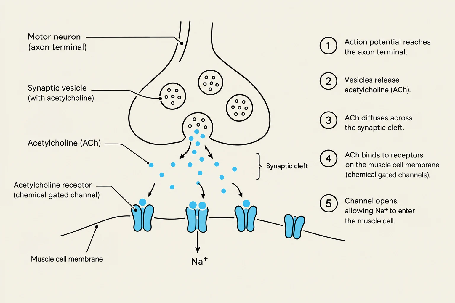
When the impulse reaches the end of the neuron, it triggers the release of a chemical neurotransmitter called acetylcholine. This chemical travels across the tiny gap and binds to receptors on the motor end plate, a specialized region of the muscle cell membrane. These receptors are actually chemical gated ion channels. Once acetylcholine binds to them, the gates open, allowing ions to flow into the cell. This creates a local change in the membrane potential—a "graded potential"—which, if strong enough, triggers a full-scale electrical impulse in the muscle fiber.
Diagram illustrating the process of synaptic transmission at a neuromuscular junction, showing the release of acetylcholine from a motor neuron and its binding to receptors on a muscle cell membrane, leading to the opening of ion channels and muscle cell excitation.
The Signal: Action Potentials and Propagation
Once the motor end plate reaches a specific electrical "threshold," it triggers an action potential. An action potential is a brief, temporary change in the membrane potential where the cell moves from a negative resting state to a positive state, and then back again.
This electrical wave is driven by two main types of voltage gated channels:
Sodium (Na+) Channels: When the threshold is reached, these channels open rapidly, allowing sodium to rush into the cell (influx). This causes depolarization, making the inside of the cell much more positive.
Potassium ($K^+$) Channels: As the cell reaches its maximum positive potential, these channels open to allow potassium to exit the cell (efflux). This causes repolarization, bringing the membrane potential back down toward its resting state.
Because these channels open in response to the voltage changes caused by their neighbors, the action potential moves like a falling row of dominoes along the entire length of the muscle fiber.
Exam Tip: Remember the difference between "influx" and "efflux." Influx means ions are moving into the cell (like Sodium during depolarization), while efflux means they are moving out of the cell (like Potassium during repolarization).
The Bridge: T-Tubules and the Triad
A muscle fiber is a large cell, and a signal on the surface might not reach the deep interior quickly enough to cause a synchronized contraction. To solve this, the sarcolemma (the muscle cell membrane) features deep, continuous invaginations called T-tubules (Transverse Tubules).
The T-tubules act as "highways," carrying the action potential from the surface deep into the center of the muscle cell. Nestled against these T-tubules are the terminal cisternae, which are enlarged sacs of the sarcoplasmic reticulum (SR)—an internal organelle that acts as a massive storage tank for calcium ions.
The functional unit where one T-tubule is flanked by two terminal cisternae is called a triad. This triad is the critical "conversion station" where the electrical signal is turned into a chemical release.
Diagram illustrating the structure of a muscle fiber, including the sarcolemma, T-tubule, sarcoplasmic reticulum, terminal cisternae, and myofibrils.
The Trigger: Excitation-Contraction Coupling
Inside the membrane of the T-tubule, there are specialized voltage sensitive proteins. These proteins are physically and mechanically linked to mechanically gated ion channels located on the membrane of the sarcoplasmic reticulum.
The sequence of events within the triad is as follows:
The action potential travels down the T-tubule.
The electrical change in voltage causes the voltage sensitive protein to change its shape (conformation).
Because the protein is physically attached to the mechanically gated ion channel on the SR, this shape change pulls the gate open.
Calcium ions, which were stored at high concentrations inside the SR, flood out into the cytoplasm of the muscle cell.
The Result: Muscle Contraction
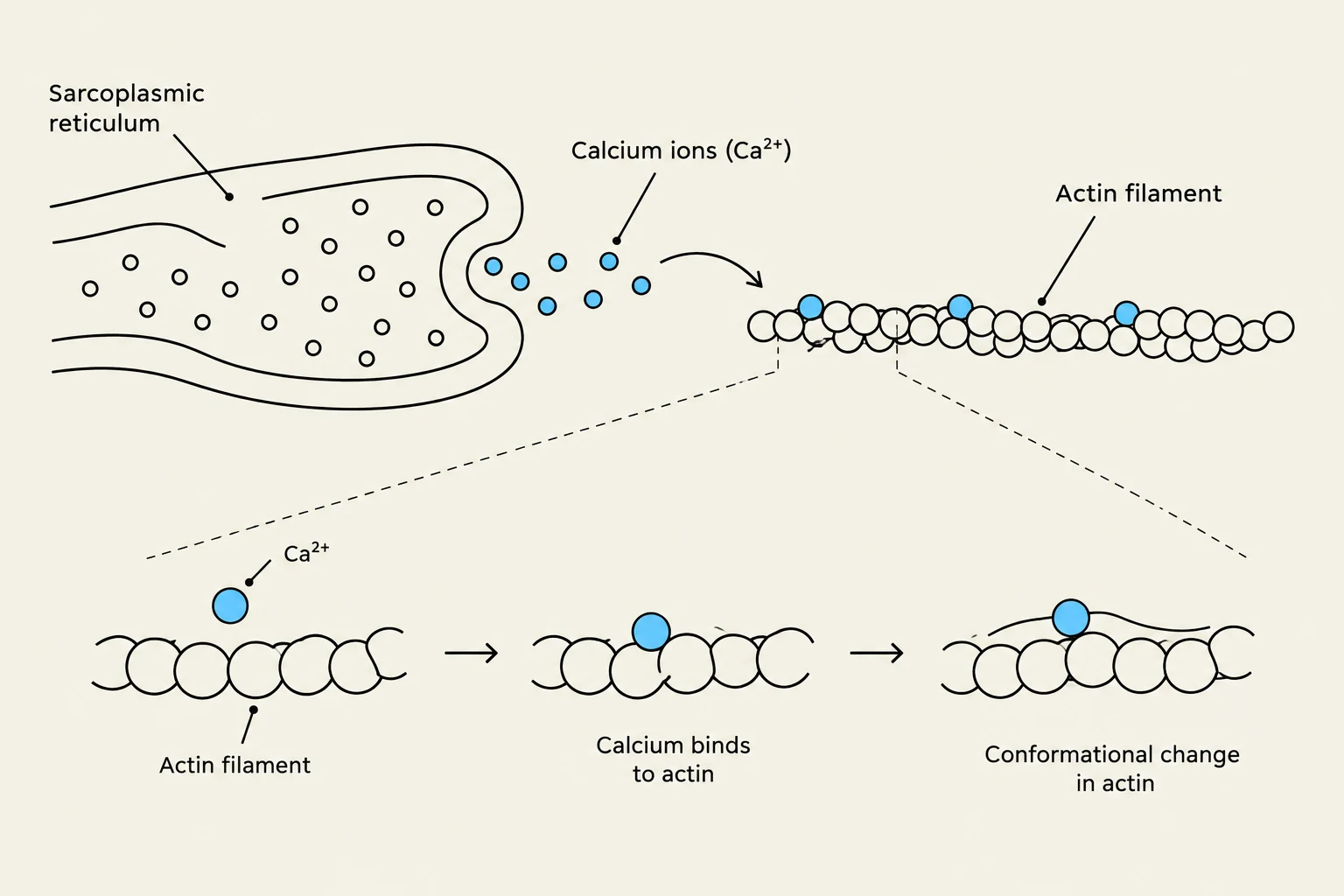
The sudden flood of calcium is the final trigger for movement. The calcium ions bind to the thin filaments, specifically the actin proteins. This binding changes the shape of the actin, allowing it to interact with other proteins to initiate the "sliding filament" mechanism. This molecular tug-of-war shortens the muscle fiber, resulting in a physical contraction.
Diagram illustrating the role of calcium ions in muscle contraction. Calcium ions released from the sarcoplasmic reticulum bind to actin filaments, causing a conformational change that facilitates muscle contraction.
Summary Table: Ion Movement and Membrane Potential
Phase
Primary Ion Movement
Channel Type
Effect on Potential
Depolarization
Na+ Influx (In)
Voltage-gated Sodium
Becomes more positive
Repolarization
$K^+$ Efflux (Out)
Voltage-gated Potassium
Returns to negative
Hyperpolarization
$K^+$ Efflux (Continued)
Voltage-gated Potassium
Becomes more negative than resting
Keep reading for free.
Create an account to finish this reading and make it part of your study routine.