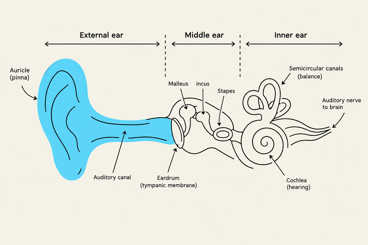
The Tympanic Membrane
As the clinician moves deeper into the ear using an otoscope, the next landmark is the tympanic membrane (eardrum). The appearance of the membrane provides significant diagnostic clues:
- Healthy Membrane: Appears pearly gray, translucent, and smooth, with a visible "cone of light" (light reflex).
- Abnormalities: A bulging, opaque, or reddish membrane may indicate middle ear effusion or infection (acute otitis media). A perforated membrane may show a visible hole or tear, sometimes containing a tympanostomy tube for pressure equalization.
Clinical Tip: Assessing Mobility
When using an otoscope with a pneumatic attachment, observe the mobility of the tympanic membrane. A lack of mobility in a bulging membrane often suggests middle ear effusion due to acute otitis media.
Evaluating Hearing and Equilibrium
The function of Cranial Nerve VIII (the Vestibulocochlear nerve) is assessed through hearing and equilibrium evaluations.
Hearing Assessment
A hearing screening should begin simply by observing the patient's ability to respond to verbal questions and directions. Clinicians should remain vigilant for compensatory behaviors that suggest impairment, such as:
- Cupping a hand behind the ear to amplify sound.
- Tilting the ear toward the examiner to improve sound localization.
More formal assessments may include the whispered voice test or the use of tuning forks (typically 512 and 1024 Hz) to test air and bone conduction.
The Nose and Sinuses
The nasal examination focuses on the patency of the airway and the health of the mucosal linings.
Inspection of the Nasal Cavity
The nasal mucosa should appear pink, moist, and glistening. Key structures to inspect include:
- Nasal Septum: This should be relatively straight and close to the midline, with the anterior portion being thicker than the posterior. A deviation may indicate asymmetric posterior nasal cavities. Note that crusting on the anterior septum (at the Kiesselbach plexus) may occur following epistaxis (nosebleed).
- Turbinates: These should be firm and the same color as the surrounding tissue. A bluish-gray or pink color with a "boggy" (swollen) consistency is often indicative of allergies.
- Discharge: While moist, clear discharge on the septum is common, turbinate discharge may signal sinusitis, an upper respiratory infection, or a foreign body.
Abnormal Findings
Clinicians must look for masses or localized swelling. For example, a polyp presents as a rounded, elongated mass projecting from the boggy mucosa into the cavity. Additionally, chronic cocaine use can lead to the destruction of the nasal mucosa and perforation of the septum.
The Special Senses: Smell and Taste
The examination of the ENT region is inextricably linked to the special senses.
- Smell: This sense is mediated by Cranial Nerve I (the Olfactory nerve). It is typically assessed by asking the patient to recognize various familiar odors.
- Taste: While often assessed alongside the mouth and throat examination, taste is a critical sensory function associated with the oral cavity and the integrity of the cranial nerves.
Clinical Considerations Across the Lifespan
The findings of an ENT exam must be interpreted within the context of the patient's age and history:
- Children: Be alert for language delays or a lack of reaction to names, which may indicate hearing loss. History of recurrent otitis media is also common.
- Older Adults: Hearing loss is common and can interfere with daily life. Additionally, many medications used by older adults (such as diuretics or antihypertensives) can cause dry mouth (xerostomia).
- Environmental Factors: History of exposure to loud noises (factories, headphones) is a significant risk factor for hearing loss.