Eukaryotic Cell Division: Mitosis and the Cell Cycle
An in-depth explanation of the eukaryotic cell cycle, detailing interphase, mitosis stages (prophase, prometaphase, metaphase, anaphase, telophase), cytokinesis, chromosomal organization, karyotypes, and cell cycle control mechanisms including checkpoints and their relation to cancer.
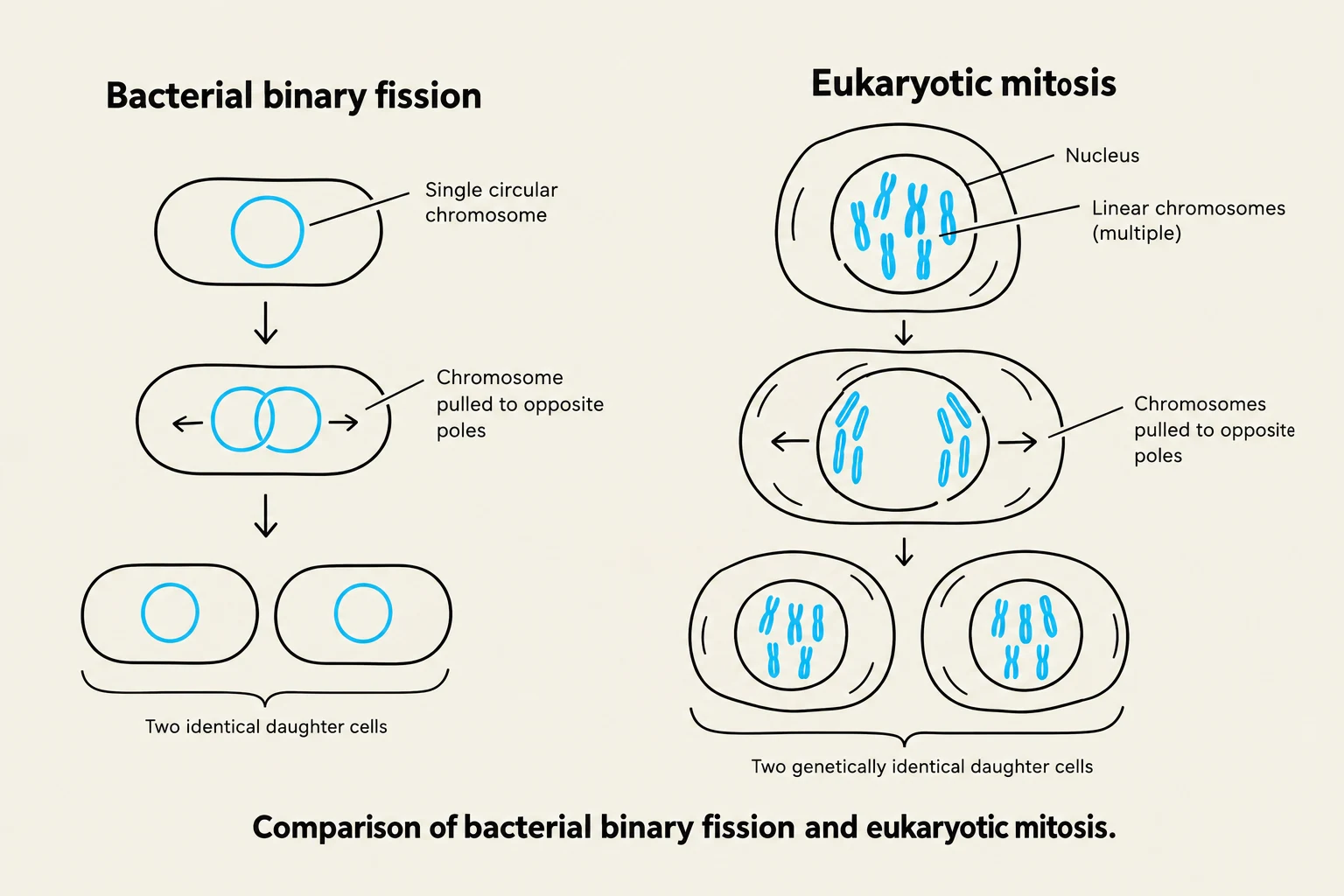
Cell division is the fundamental process by which life propagates, ensuring that genetic information is accurately passed from one generation of cells to the next. While all living organisms must divide to survive, the complexity of the process varies significantly between domains. While bacteria undergo a relatively simple process of binary fission—where a single, circular chromosome is replicated and partitioned to opposite ends of the cell—eukaryotic cells must manage much larger, linear chromosomes organized within a complex nucleus.
Comparison of bacterial binary fission and eukaryotic mitosis.
To prevent errors during this complex dance, eukaryotic cells utilize a highly regulated cycle of growth, DNA replication, and division.
The Eukaryotic Cell Cycle
The eukaryotic cell cycle is a continuous process divided into two main stages: interphase and mitosis, followed by cytokinesis.
Interphase: Preparation for Division
Interphase is the longest part of the cell cycle, during which the cell grows and prepares its molecular machinery for division. It is subdivided into three distinct phases:
G1 phase (Gap 1): This is the primary growth phase where the cell increases in size and performs its normal metabolic functions.
S phase (Synthesis): This is the critical stage of DNA replication. Before this phase, each chromosome consists of a single DNA molecule. After replication, each chromosome is composed of two identical sister chromatids held together by cohesin proteins.
G2 phase (Gap 2): The cell continues to grow, organelles replicate, and chromosomes begin to coil more tightly using motor proteins to prepare for the physical rigors of mitosis.
Mitosis and Cytokinesis
Mitosis is the process of nuclear division, ensuring each daughter cell receives an identical set of chromosomes. Once the nucleus has divided, cytokinesis physically splits the cytoplasm into two new cells.
In animal cells: A cleavage furrow is formed by the contraction of actin filaments, pinching the cell in two.
In plant cells: Because of the rigid cell wall, a cell plate forms between the two new nuclei to create a new partition.
The Five Stages of Mitosis
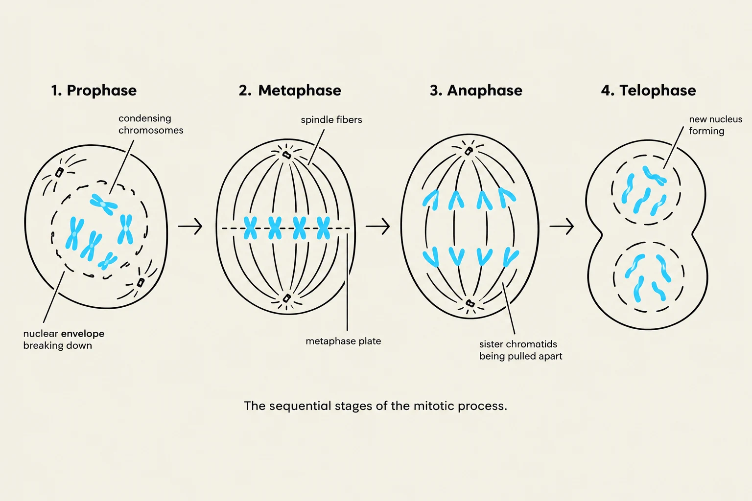
The sequential stages of the mitotic process, illustrating Prophase, Metaphase, Anaphase, and Telophase.
Mitosis is a highly choreographed sequence of events that ensures accurate chromosome segregation.
Prophase: Chromosomes condense and become visible under a light microscope. The nuclear envelope begins to break down, and the spindle apparatus starts to assemble.
Prometaphase: The nuclear envelope is fully disassembled. Microtubules from the spindle apparatus attach to kinetochores, which are specialized protein structures located at the centromere (the point of constriction) of each sister chromatid.
Metaphase: The spindle fibers pull the chromosomes until they are aligned along the metaphase plate, an imaginary plane that serves as the future axis of cell division.
Anaphase: This is the stage of active separation. The cohesin proteins are removed, allowing sister chromatids to be pulled toward opposite poles. This occurs via two mechanisms:
Anaphase A: Chromatids are pulled by the shortening of kinetochore microtubules.
Anaphase B: The spindle poles themselves are pushed apart by polar microtubules.
Telophase: The chromosomes reach the poles and begin to uncoil. The nuclear envelope reforms around each set of chromosomes, and the individual sister chromatids are now considered individual chromosomes.
Exam Tip: Mitosis vs. Cytokinesis
Remember that mitosis refers specifically to the division of the nucleus (the genetic material), whereas cytokinesis refers to the division of the cytoplasm (the cell body). They are distinct processes that occur sequentially.
Chromosomal Organization and Karyotypes
To understand how cells divide, we must understand what is being divided. Chromosomes are composed of chromatin, a dense complex of negatively charged DNA and positively charged proteins (histones).
Homologous Chromosomes and Ploidy
Humans are diploid (2n=46), meaning our somatic cells contain two complete sets of chromosomes. These exist as 23 pairs of homologous chromosomes. In each pair, one chromosome is inherited from the mother and one from the father. While they carry the same genes in the same order, they may contain different versions (alleles) of those genes.
Karyotypes
A karyotype is a visual representation of an individual's full set of chromosomes, arranged by size, shape, and centromere location.
Normal Human Karyotype: 46 total chromosomes (23 pairs).
Aneuploidy: Variations in chromosome number, such as having an extra or missing chromosome, are typically fatal or lead to significant developmental disorders.
Cell Cycle Control and Cancer
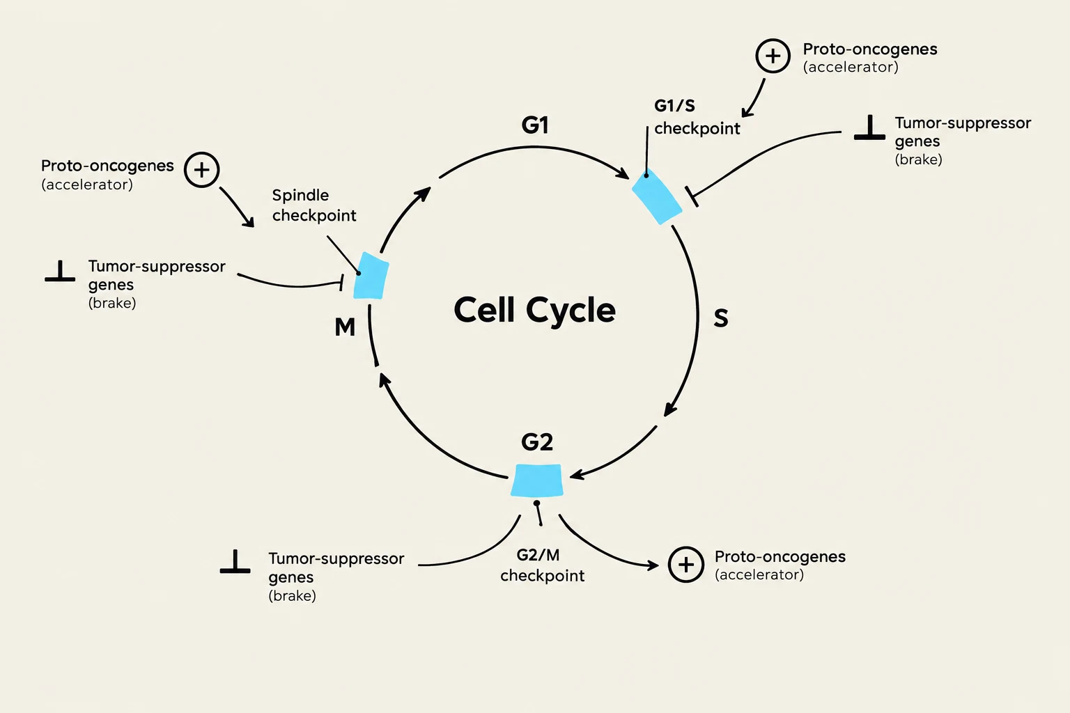
The cell cycle does not run unchecked; it is governed by a sophisticated system of checkpoints that ensure accuracy.
G1/S Checkpoint: The cell decides whether to commit to DNA replication.
G2/M Checkpoint: The cell assesses whether DNA replication was successful and if the chromosomes are ready for division.
Spindle Checkpoint: Occurs during metaphase to ensure all chromosomes are properly attached to the spindle before anaphase begins.
The primary drivers of this control are proteins known as Cyclin-Dependent Kinases (CDKs). CDKs are activated by proteins called cyclins.
When Control Fails: Cancer
Diagram illustrating the cell cycle, including checkpoints and the roles of proto-oncogenes and tumor-suppressor genes.
Cancer is essentially the result of uncontrolled cell division caused by mutations in genes that regulate the cell cycle. Two main types of genes are involved:
Tumor-suppressor genes: These act like "brakes." They monitor DNA integrity and can halt the cycle to allow for repairs. If they are mutated (e.g., the $p53$ gene), the cell may continue to divide even with damaged DNA.
Proto-oncogenes: These act like "gas pedals," promoting cell growth and division. When mutated into oncogenes, they become hyperactive, signaling the cell to divide constantly even in the absence of growth factors.
Clinical Connection: BRCA Genes
Mutations in tumor-suppressor genes like $BRCA1$ and $BRCA2$ significantly increase the risk of breast and ovarian cancers because the cell loses its ability to effectively repair DNA damage.
Keep reading for free.
Create an account to finish this reading and make it part of your study routine.